Copyright
©2013 Baishideng Publishing Group Co.
World J Gastroenterol. Jun 7, 2013; 19(21): 3354-3357
Published online Jun 7, 2013. doi: 10.3748/wjg.v19.i21.3354
Published online Jun 7, 2013. doi: 10.3748/wjg.v19.i21.3354
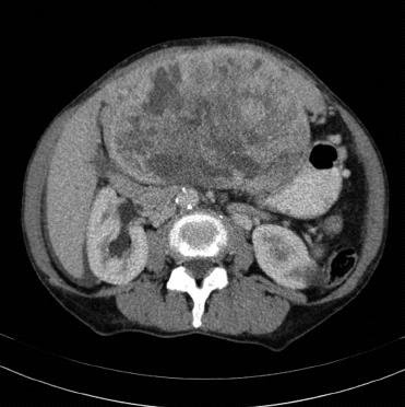
Figure 1 Computed tomography scan of the abdomen showing a well-circumscribed, solitary, large mass (15 cm × 10 cm × 20 cm) adjacent to the left liver lobe.
The claw sign of hepatic tissue suggests intrahepatic localization. Notable features include hypervascularity, heterogeneous enhancement, and smooth margins.
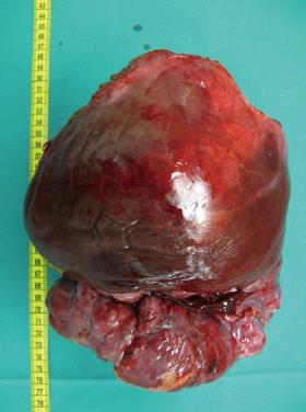
Figure 2 Gross specimen is a bulky neoplasm attached to the liver capsule.
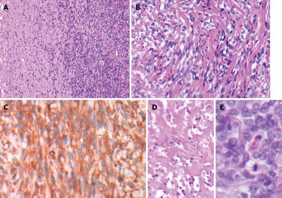
Figure 3 Histological findings indicative of malignant solitary fibrous tumor.
A: Hypo- and hypercellular patternless growth patterns [hematoxylin and eosin (HE), × 40]; B: Short spindly to ovoid tumor cells in a collagenous background (HE, × 200); C: Immunohistochemical staining with positive detection of CD34 (HE, × 200); D: Multifocal areas of necrosis (HE, × 200); E: Hypercellularity, cytologic atypia, and increased mitotic activity (HE, × 400).
- Citation: Jakob M, Schneider M, Hoeller I, Laffer U, Kaderli R. Malignant solitary fibrous tumor involving the liver. World J Gastroenterol 2013; 19(21): 3354-3357
- URL: https://www.wjgnet.com/1007-9327/full/v19/i21/3354.htm
- DOI: https://dx.doi.org/10.3748/wjg.v19.i21.3354


