Copyright
©2013 Baishideng Publishing Group Co.
World J Gastroenterol. Jun 7, 2013; 19(21): 3352-3353
Published online Jun 7, 2013. doi: 10.3748/wjg.v19.i21.3352
Published online Jun 7, 2013. doi: 10.3748/wjg.v19.i21.3352
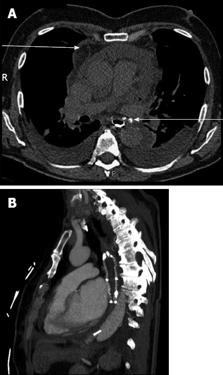
Figure 1 Computed tomography-scan.
A: Computed tomography (CT)-scan with esophageal opacification. Pericardial effusion (upper arrow) and esophagopericardial fistula (lower arrow) were both present; B: CT-scan performed after esophageal stent placement and surgical drainage. Pericardial effusion was no longer present following esophageal stent insertion.
- Citation: Quénéhervé L, Musquer N, Léauté F, Coron E. Endoscopic management of an esophagopericardial fistula after radiofrequency ablation for atrial fibrillation. World J Gastroenterol 2013; 19(21): 3352-3353
- URL: https://www.wjgnet.com/1007-9327/full/v19/i21/3352.htm
- DOI: https://dx.doi.org/10.3748/wjg.v19.i21.3352


