Copyright
©2013 Baishideng Publishing Group Co.
World J Gastroenterol. Jun 7, 2013; 19(21): 3173-3188
Published online Jun 7, 2013. doi: 10.3748/wjg.v19.i21.3173
Published online Jun 7, 2013. doi: 10.3748/wjg.v19.i21.3173
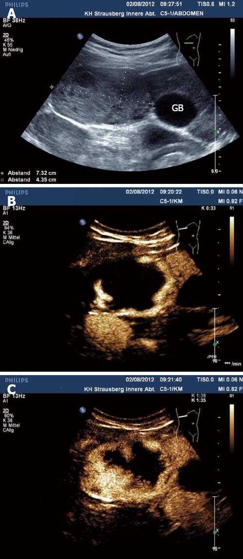
Figure 1 Hemangioma.
Large hemangioma in B-mode (A), with typical peripheral nodular contrast enhancement (B) and centripetal fill-in (C). GB: Gallbladder.
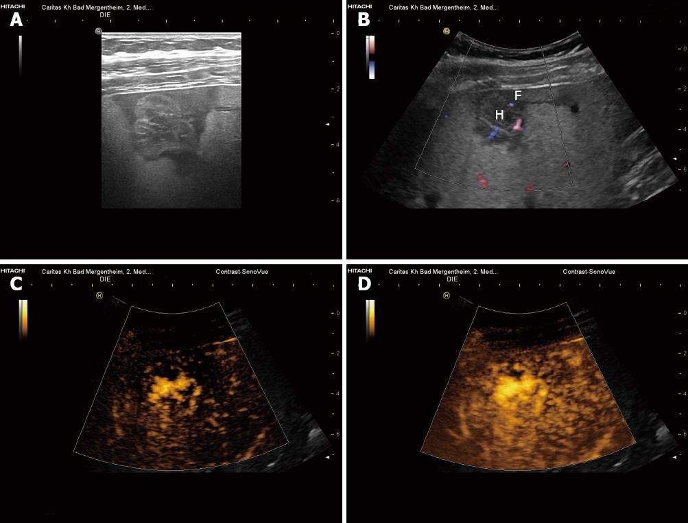
Figure 2 Shunt hemangioma.
Shunt hemangiomas are typically small (often < 20 mm) with abundant arterio(porto-)venous shunts (functionally described as high flow hemangiomas). They are often surrounded by less fat-containing hypoechoic liver parenchyma (A, B) due to the dominant arterial blood flow in comparison to the reduced portal venous perfusion. Arterial contrast enhancement of the shunt-hemangioma is also shown (C, D). H: Hemangioma; F: Less fat-containing hypoechoic area. Reproduced with permission from Dietrich et al[65].
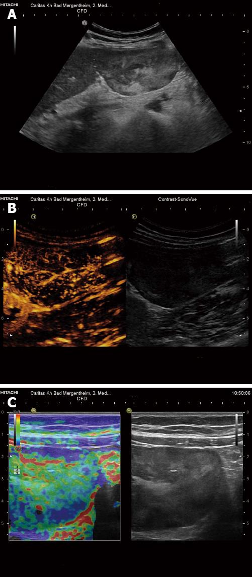
Figure 3 Teleangiectatic focal nodular hyperplasia.
Pedunculated liver tumor, histologically teleangiectatic focal nodular hyperplasia with signs of peliosis. A: B-mode imaging shows heterogenous echogenicity; B: Contrast-enhanced ultrasound reveals central arterial blood supply; C: Real-time elastography shows harder periphery and softer central portions of the lesion.
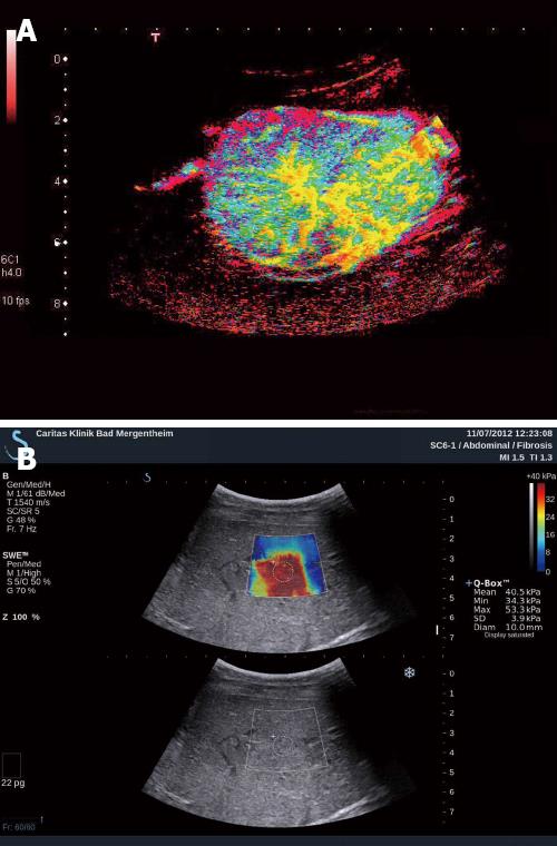
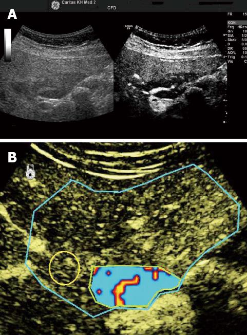
Figure 4 Focal nodular hyperplasia.
A: In addition to conventional contrast-enhanced ultrasound (CEUS), parametric CEUS displays also the timeline of contrast enhancement (early enhancement in yellow, later enhancement in blue); B: Histologically proven. B-mode revealed isoechoic lesion, very difficult to identify. Shear wave elastography reveals very hard tissue of the lesion, shown in red in comparison to the surrounding soft liver parenchyma.
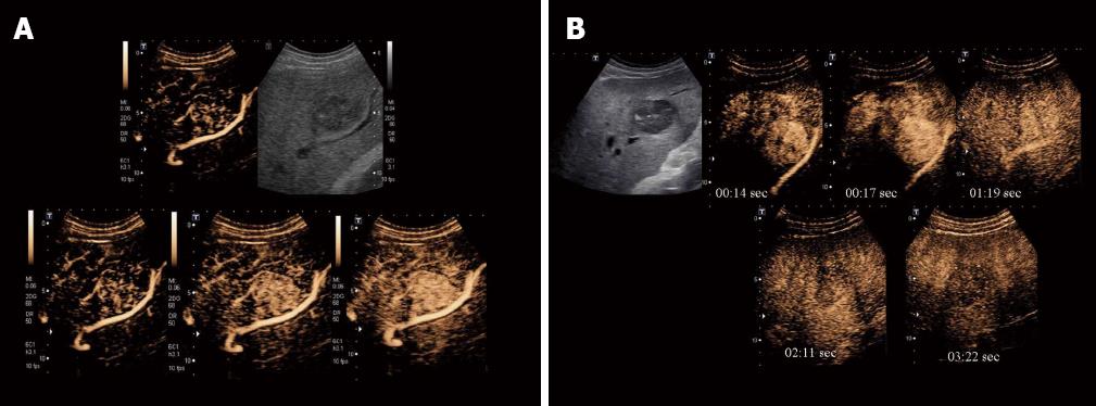
Figure 5 Focal fatty sparing.
A: Focal fatty changes may simulate masses on conventional B-mode ultrasound; B: In the arterial, portal venous and late phases, focal fatty changes show similar enhancement patterns to that of the adjacent liver parenchyma. Contrast-enhanced ultrasound is helpful for the identification of the centrally located arteries. Typically centrally located arteries (and often also portal venous branches and hepatic veins) can be identified. Dynamic vascular pattern improves contrast imaging. Reproduced with permission from Cui et al[120].
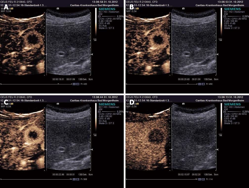
Figure 6 Liver metastasis.
In contrast to the variably enhancing arterial phase (A-C), liver metastases are typically hypoenhancing during the portal venous (sinusoidal) phase (D) which facilitates their reliable diagnosis.
Figure 7 Hepatocellular carcinoma with arterial hyperenhancement (A) and hypoenhancement in the portal venous phase (B).
- Citation: Dietrich CF, Sharma M, Gibson RN, Schreiber-Dietrich D, Jenssen C. Fortuitously discovered liver lesions. World J Gastroenterol 2013; 19(21): 3173-3188
- URL: https://www.wjgnet.com/1007-9327/full/v19/i21/3173.htm
- DOI: https://dx.doi.org/10.3748/wjg.v19.i21.3173


