Copyright
©2013 Baishideng Publishing Group Co.
World J Gastroenterol. May 28, 2013; 19(20): 3169-3172
Published online May 28, 2013. doi: 10.3748/wjg.v19.i20.3169
Published online May 28, 2013. doi: 10.3748/wjg.v19.i20.3169
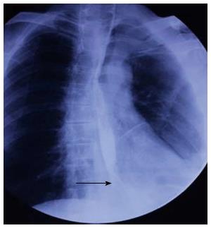
Figure 1 Barium esophagography.
Esophagography shows a type 1 tumor, 1.5 cm in length, in the lower thoracic esophagus (arrow).
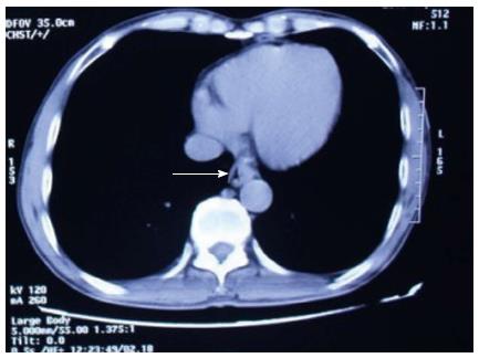
Figure 2 Thoracic computed tomography shows a tumor 1 cm in diameter (arrow).
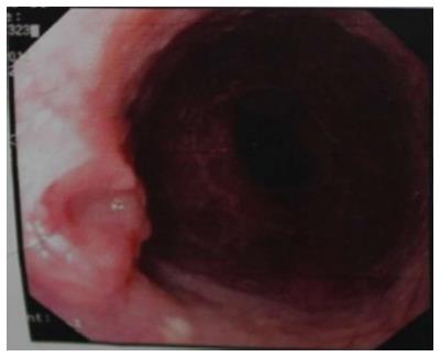
Figure 3 Upper gastrointestinal endoscopy shows a tumor 39 cm from the incisor, the anterior wall of the esophagus has a lip bulge about 1.
2 cm × 1.0 cm.
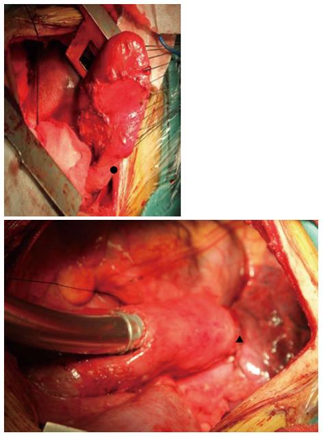
Figure 4 Esophagus is anastomosed to the remnant stomach.
During surgery, after cutting the left, right and short gastric vessels, the remnant stomach relies on the blood supply of the anastomotic stoma alone and is still rosy. Circle: Jejunum-stomach anastomotic stoma; Triangle: Esophagus-remnant stomach anastomotic stoma.
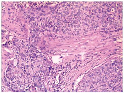
Figure 5 The tumor was diagnosed as highly differentiated squamous cell carcinoma.
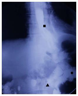
Figure 6 Barium esophagography on the 7th postoperative day.
Esophagography shows that barium passes smoothly through the anastomotic stoma, without leakage. Square: Anastomotic stoma; Triangle: Afferent loop of jejunum; Circle: Efferent loop of jejunum.
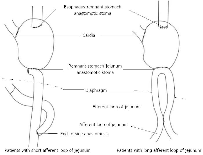
Figure 7 Scheme of the resection area of the gastric remnant and the esophagus (pre-operative), and reconstruction of the organs (post-operative).
- Citation: Xie SP, Fan GH, Kang GJ, Geng Q, Huang J, Cheng BC. Esophageal reconstruction with remnant stomach: A case report and review of literature. World J Gastroenterol 2013; 19(20): 3169-3172
- URL: https://www.wjgnet.com/1007-9327/full/v19/i20/3169.htm
- DOI: https://dx.doi.org/10.3748/wjg.v19.i20.3169


