Copyright
©2013 Baishideng Publishing Group Co.
World J Gastroenterol. May 14, 2013; 19(18): 2781-2792
Published online May 14, 2013. doi: 10.3748/wjg.v19.i18.2781
Published online May 14, 2013. doi: 10.3748/wjg.v19.i18.2781
Figure 1 Analysis of expression of mitogen-activated protein kinase activator with WD40 repeats binding protein and mitogen-activated protein kinase activator with WD40 repeats in six gastric cancer cell lines.
A: mRNA expression for mitogen-activated protein kinase activator with WD40 repeats (MAWD)/MAWD binding protein (MAWBP) in BGC823, MGC803, SGC7901, AGS, N87 and MKN45 six gastric cancer cell lines was detected by reverse transcription-polymerase chain reaction (PCR). There was a low level of endogenous MAWBP and MAWD mRNA in SGC7901 cells, and a high level in BGC823 cells; B: Expression of MAWBP and MAWD proteins was detected by Western blotting. The expression level was lower in SGC7901 cells than in the other cell lines. Expression of MAWBP and MAWD was higher in BGC823 cells than in the other cells; C: mRNA expression for MAWBP and MAWD was detected by real-time PCR. There were lower levels of endogenous MAWD and MAWBP mRNA in SGC7901 cells, and higher levels in BGC823 cells. NTC: No template control.
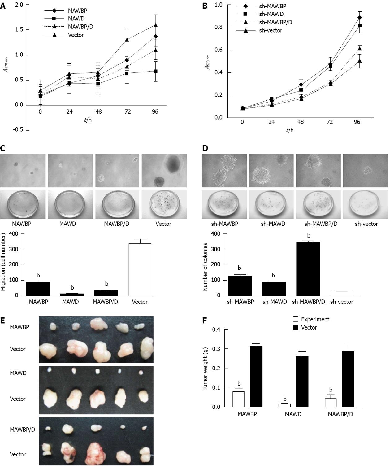
Figure 2 Effect of mitogen-activated protein kinase activator with WD40 repeats binding protein and mitogen-activated protein kinase activator with WD40 repeats on proliferation and tumorigenicity of gastric cancer cells.
A: 3-(4,5-Dimethyl-2-thiazolyl)-2,5-diphenyl tetrazolium bromide assay showed that growth of SGC7901 cells overexpressing mitogen-activated protein kinase activator with WD40 repeats (MAWD)/MAWD binding protein (MAWBP) was markedly inhibited; B: In BGC823 cells with inhibition of expression of MAWBP and MAWD, cell growth was inversely increased; C: In soft agar assay, colonies of cells with overexpression of MAWBP, MAWD and MAWBP/D were reduced in number and size compared with cells transfected with vectors alone (original magnification of clones: ×100); D: Knockdown group demonstrated the opposite effects. There was an increase in the number and size of sh-MAWBP, sh-MAWD, sh-MAWBP/D cells compared with cells transfected with vectors alone; E: Nude mouse xenografts. Tumors induced by MAWBP, MAWD and MAWBP/D cotransfected cells were smaller than those of the vector group; F: Xenograft weight. The average tumor weight were calculated from five nude mice in every group. Data are presented as the mean ± SD from three independent experiments. bP < 0.01 vs vector group.
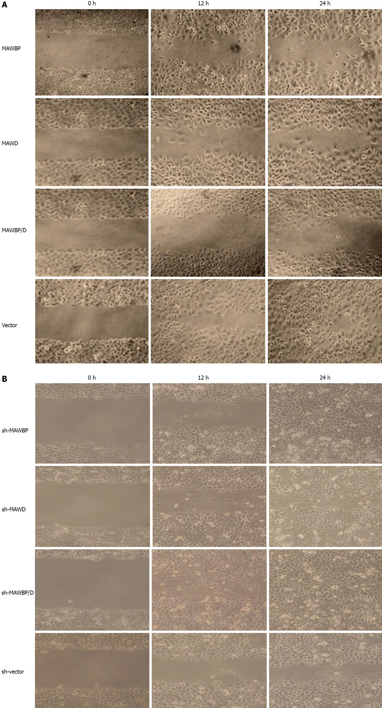
Figure 3 Effect of mitogen-activated protein kinase activator with WD40 repeats binding protein and mitogen-activated protein kinase activator with WD40 repeats on migration of gastric cancer cells.
A: Wound healing assay showed that migration of vector-transfected cells was faster than that of cells overexpressing mitogen-activated protein kinase activator with WD40 repeats (MAWD)/MAWD binding protein (MAWBP). The scratch gap in vector group was almost closed at 24 h. The migration of MAWBP/D cotransfected cells was slowest; B: Cells with combined downregulation of MAWBP and MAWD expression migrated faster than the other cells. Migration of sh-vector cells was slowest (original magnification, ×100).
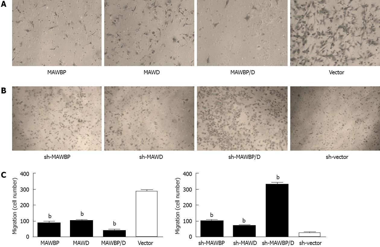
Figure 4 Effect of mitogen-activated protein kinase activator with WD40 repeats binding protein and mitogen-activated protein kinase activator with WD40 repeats on invasive ability of gastric cancer cells.
A: Transwell assay showed that invasive ability of mitogen-activated protein kinase activator (MAWD) with WD40 repeats binding protein (MAWBP)/D cotransfected cells was weakest, with the lowest number of cells to cross the matrix membranes. Vector-transfected cells migrated farthest; B: Knockdown of MAWBP and MAWD increased the invasive ability of gastric cancer (GC) cells (original magnification: ×100); C: Number of cells that traverse the matrix membrane in the different groups. Data are presented as the mean ± SD from three independent experiments. bP < 0.01 vs vector group.
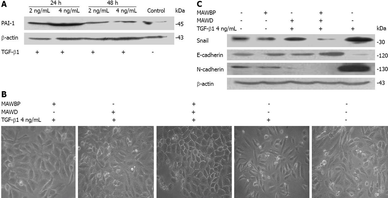
Figure 5 Effect of mitogen-activated protein kinase activator with WD40 repeats binding protein and mitogen-activated protein kinase activator with WD40 repeats on expression of biomarkers specific for epithelial-mesenchymal transition induced by transforming growth factor-β1.
A: According to the expression of transforming growth factor (TGF)-β downstream reporter gene plasminogen activator inhibitor (PAI)-1, the optimum TGF-β1 concentration and time were confirmed as 4 ng/mL for 24 h; B: SGC7901 cells overexpressing mitogen-activated protein kinase activator with WD40 repeats (MAWD)/MAWD binding protein (MAWBP) were stimulated by 4 ng/mL TGF-β1 for 24 h. Cells that overexpressed both MAWBP and MAWD displayed a pebble-like shape, while vector cells showed a classical mesenchymal phenotype (original magnification, ×400); C: Expression of E-cadherin was strongest in the MAWBP/D cotransfection group and weakest in the vector group, using Western blotting. Snail and N-cadherin were inversely associated with E-cadherin expression.
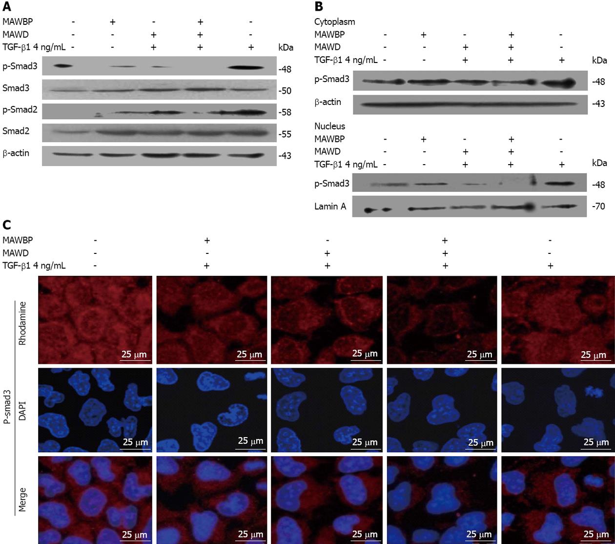
Figure 6 Combination of mitogen-activated protein kinase activator with WD40 repeats binding protein and mitogen-activated protein kinase activator with WD40 repeats inhibited the transforming growth factor-β pathway.
A: The level of p-Smad3 was lowest in the mitogen-activated protein kinase activator (MAWD) with WD40 repeats binding protein (MAWBP)/D cotransfected group and highest in the vector group. p-Smad2 was also lower in the MAWBP/D group; B, C: Nuclear translocation capability of p-Smad3 in cotransfected cells was weakest that means the activity of transforming growth factor (TGF)-β pathway in co-expression group was inhibited. DAPI: 4’,6-diamidino-2-phenylindole.
- Citation: Li DM, Zhang J, Li WM, Cui JT, Pan YM, Liu SQ, Xing R, Lu YY. MAWBP and MAWD inhibit proliferation and invasion in gastric cancer. World J Gastroenterol 2013; 19(18): 2781-2792
- URL: https://www.wjgnet.com/1007-9327/full/v19/i18/2781.htm
- DOI: https://dx.doi.org/10.3748/wjg.v19.i18.2781


