Copyright
©2013 Baishideng Publishing Group Co.
World J Gastroenterol. May 7, 2013; 19(17): 2714-2717
Published online May 7, 2013. doi: 10.3748/wjg.v19.i17.2714
Published online May 7, 2013. doi: 10.3748/wjg.v19.i17.2714
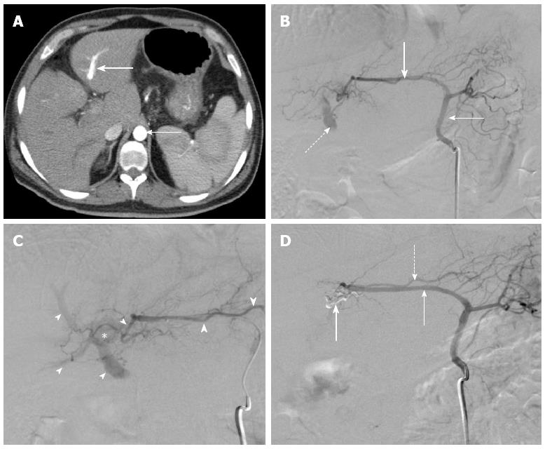
Figure 1 Intrahepatic communication between hepatic artery and portal vein.
A: Contrast enhanced axial computed tomography of the abdomen demonstrating similar contrast opacification of the aorta (thin arrow) and the left portal vein (thick arrow); B: Left gastric arteriogram demonstrates gastrohepatic trunk (thin arrow) giving rise to aberrant left hepatic artery (thick arrow). Note early opacification of the portal vein (dashed arrow); C: Selective arteriogram with coaxial microcatheter in the left hepatic artery demonstrates medial branch of the left hepatic artery (large arrowheads) contributing to a parenchymal blush (star) and leading to opacification of the portal veins (small arrowheads). Note the normal appearance of the lateral branch of the left hepatic artery (thick arrow); D: Post-embolization arteriogram in the gastrohepatic trunk demonstrates opacification of the left hepatic artery (thin arrow). Onyx cast (thick arrow) is seen occupying the previously seen medial branch of the left hepatic artery. No further opacification of the portal veins is seen. Note the preserved lateral branch of the left hepatic artery (dashed arrow).
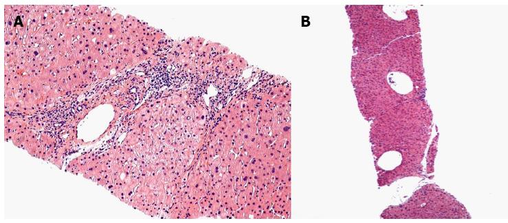
Figure 2 The hepatic histology of arterioportal fistula.
A: This low power image shows a small portal tract in the center of the field that has a markedly dilated portal venule. The terminal hepatic (central) venules above and below the portal tract are markedly dilated; B: The portal venule is slightly dilated and there are increased numbers of small portal venous radicals.
- Citation: Nookala A, Saberi B, Ter-Oganesyan R, Kanel G, Duong P, Saito T. Isolated arterioportal fistula presenting with variceal hemorrhage. World J Gastroenterol 2013; 19(17): 2714-2717
- URL: https://www.wjgnet.com/1007-9327/full/v19/i17/2714.htm
- DOI: https://dx.doi.org/10.3748/wjg.v19.i17.2714


