Copyright
©2013 Baishideng Publishing Group Co.
World J Gastroenterol. May 7, 2013; 19(17): 2668-2675
Published online May 7, 2013. doi: 10.3748/wjg.v19.i17.2668
Published online May 7, 2013. doi: 10.3748/wjg.v19.i17.2668
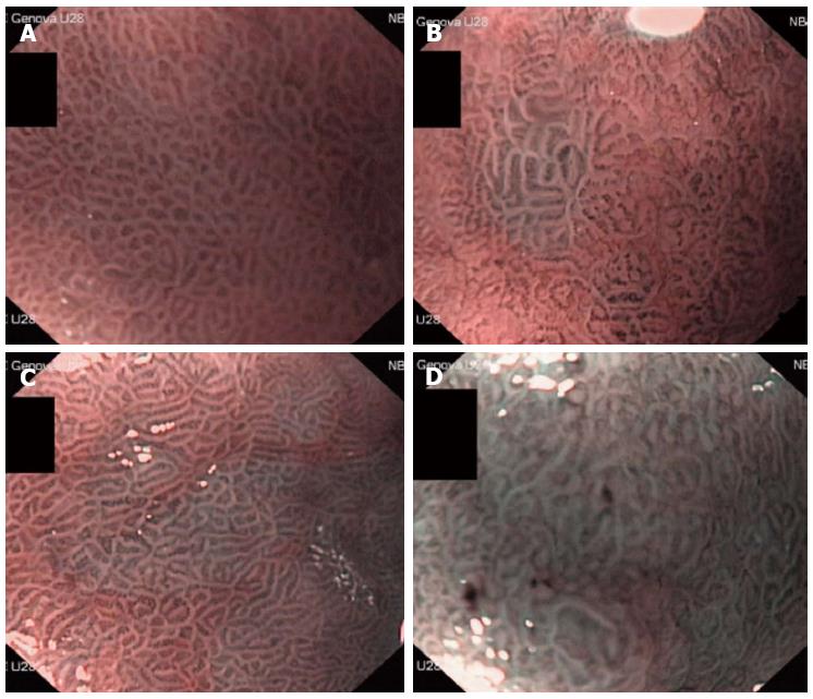
Figure 1 Normal mucosal aspects.
A: Narrow-band-imaging with magnification endoscopy examination and the degree of light blue crest appearance; B: Light blue crest (LBC) +, less than 20%; C: LBC ++, 20%-80%; D: LBC +++, 80% or more.
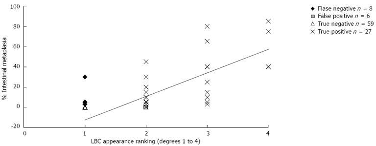
Figure 2 Graphic representation of the correlation between the degree of light blue crest appearance during narrow-band imaging with magnifying endoscopy examination and the percentage of gastric intestinal metaplasia detected by histological examination.
LBC: Light blue crest.
- Citation: Savarino E, Corbo M, Dulbecco P, Gemignani L, Giambruno E, Mastracci L, Grillo F, Savarino V. Narrow-band imaging with magnifying endoscopy is accurate for detecting gastric intestinal metaplasia. World J Gastroenterol 2013; 19(17): 2668-2675
- URL: https://www.wjgnet.com/1007-9327/full/v19/i17/2668.htm
- DOI: https://dx.doi.org/10.3748/wjg.v19.i17.2668


