Copyright
©2013 Baishideng Publishing Group Co.
World J Gastroenterol. Apr 28, 2013; 19(16): 2578-2582
Published online Apr 28, 2013. doi: 10.3748/wjg.v19.i16.2578
Published online Apr 28, 2013. doi: 10.3748/wjg.v19.i16.2578
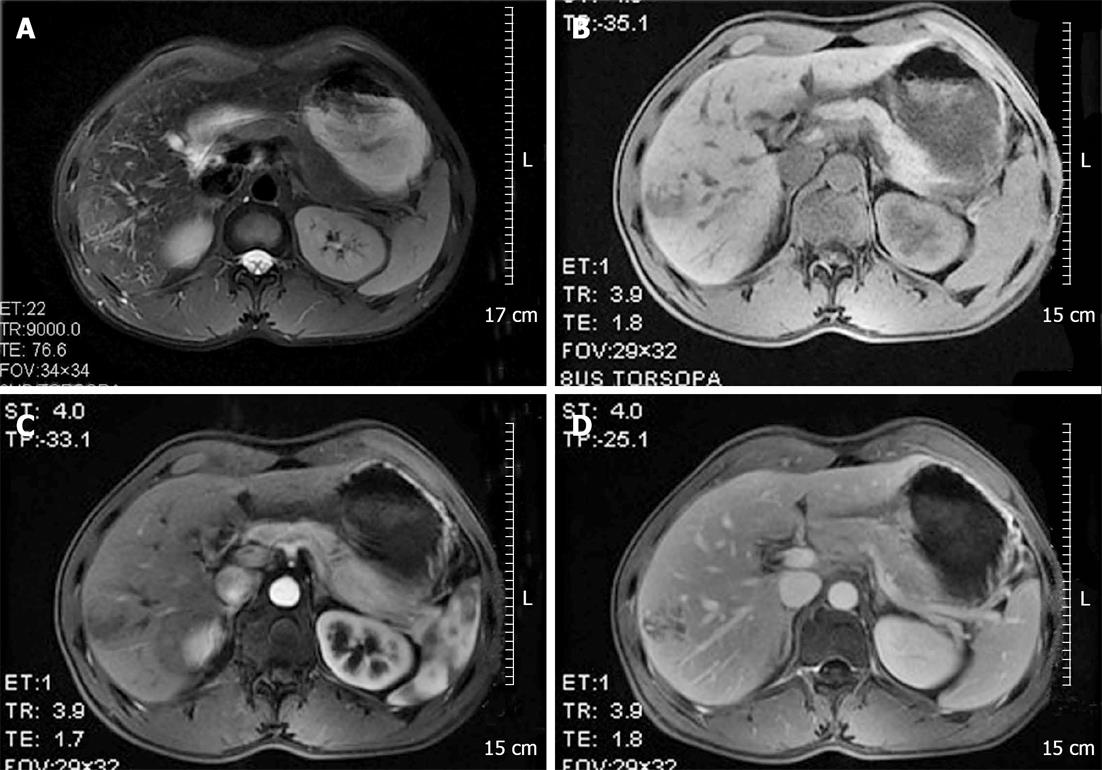
Figure 1 Magnetic resonance imaging scans.
A: Hepatobiliary magnetic resonance imaging scans revealed a 3.8 cm × 3.1 cm mass in the anterior segment of the right liver with irregular boundaries, producing low-intensity T1 signals and high-intensity T2 signals; B-D: Gadolinium diethylenetriamine-pentaacid enhanced scanning showed inhomogeneous enhancement, peaking during the arterial phase and declining during the portal vein and parenchymal phases. The lumen and collateral vessels of the portal vein were well defined without filling defects.
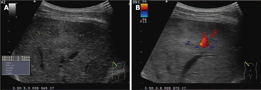
Figure 2 B-mode ultrasonography.
A: B-mode ultrasonography showing a 3.9 cm × 3.3 cm mass with inhomogeneous echo in the right hemiliver; B: The boundary was unclear and some blood-flow signal was detected within the mass.
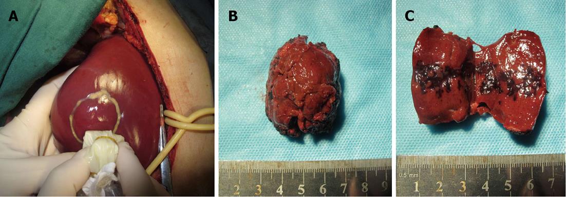
Figure 3 Gross appearance of the mass after surgical removal.
A: There were no clear boundaries or lining membrane between the mass and the liver; B: The specimen had an irregular shape; C: Several blood-filled cystic spaces were observed after opening the mass.
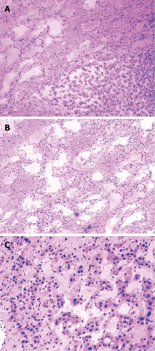
Figure 4 Pathologic examination, showing irregular and dilated lacunae, some of which were filled with blood and without endothelial linings.
Hematoxylin and eosin staining, original magnification A: × 100; B: × 200; C: × 400.
- Citation: Pan W, Hong HJ, Chen YL, Han SH, Zheng CY. Surgical treatment of a patient with peliosis hepatis: A case report. World J Gastroenterol 2013; 19(16): 2578-2582
- URL: https://www.wjgnet.com/1007-9327/full/v19/i16/2578.htm
- DOI: https://dx.doi.org/10.3748/wjg.v19.i16.2578


