Copyright
©2013 Baishideng Publishing Group Co.
World J Gastroenterol. Apr 14, 2013; 19(14): 2278-2281
Published online Apr 14, 2013. doi: 10.3748/wjg.v19.i14.2278
Published online Apr 14, 2013. doi: 10.3748/wjg.v19.i14.2278
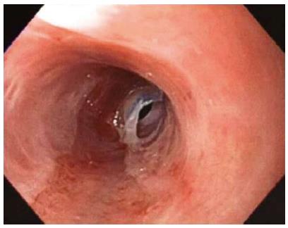
Figure 1 Endoscopy showing webs.
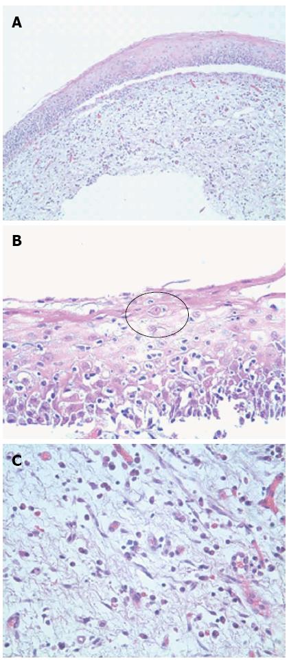
Figure 2 Histologically, the esophageal tissue demonstrated extensive denudation of the surface epithelium.
A: Subepithelial separation, HE stain, × 100; B: Civatte bodies (black circle), HE stain, × 400; C: Subepithelial edema and inflammation, HE stain, × 400.
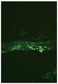
Figure 3 Immunofluorescence, fibrinogen.
- Citation: Nielsen JA, Law RM, Fiman KH, Roberts CA. Esophageal lichen planus: A case report and review of the literature. World J Gastroenterol 2013; 19(14): 2278-2281
- URL: https://www.wjgnet.com/1007-9327/full/v19/i14/2278.htm
- DOI: https://dx.doi.org/10.3748/wjg.v19.i14.2278


