Copyright
©2013 Baishideng Publishing Group Co.
World J Gastroenterol. Apr 14, 2013; 19(14): 2179-2186
Published online Apr 14, 2013. doi: 10.3748/wjg.v19.i14.2179
Published online Apr 14, 2013. doi: 10.3748/wjg.v19.i14.2179
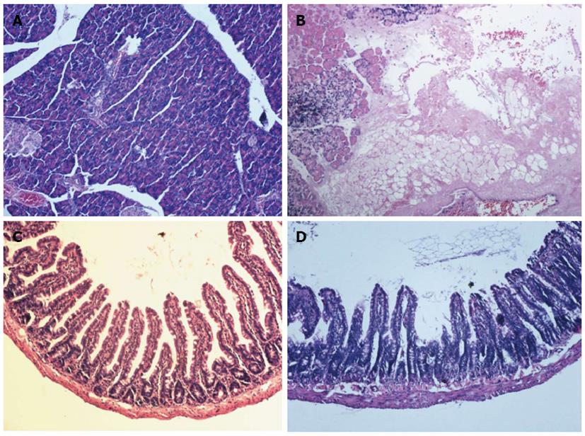
Figure 1 Histological sections from pancreas and jejunum.
A: The pancreas of the sham (S) group shows a normal exocrine and endocrine pancreatic architecture; B: The pancreas of severe acute pancreatitis (SAP) rats shows necrosis of the acinar cells accompanied by edema and hemorrhage; C: The structure of jejunum in the S group is normal; D: The mucosa was markedly denuded and the muscular layer was edematous in the SAP group. Magnification × 200.
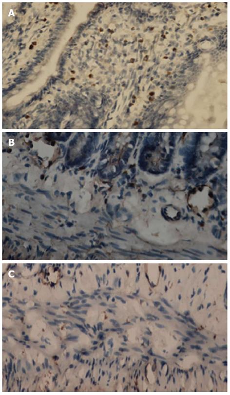
Figure 2 Immunohistochemistry for c-kit.
A: Mucosal mast cells in rats retain c-kit positivity (internal control); B: c-kit-positive interstitial cells of Cajal (ICC) in the submuscular plexus in the sham group; C: c-kit-positive ICC in the intermuscular septa in the severe acute pancreatitis group. Magnification × 200.
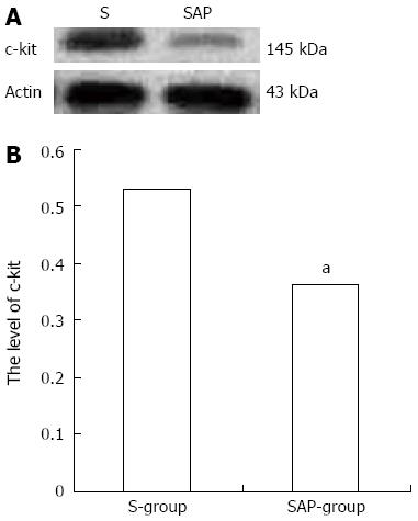
Figure 3 Expression of c-kit protein.
A: Bands of Western blotting of c-kit (145 kDa). β-actin is a loading control; B: Statistical analysis of relative density of Western blotting between two groups. Data are represented as mean ± SD. aP < 0.05 vs sham (S) group.
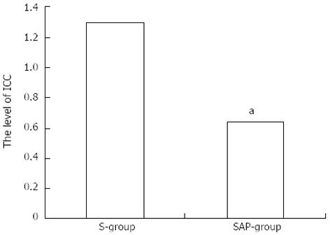
Figure 4 Mean optical density of c-kit mRNA.
Each bar represents the mean ± SD (vertical line). aP < 0.05 vs sham (S) group. ICC: Interstitial cells of Cajal.
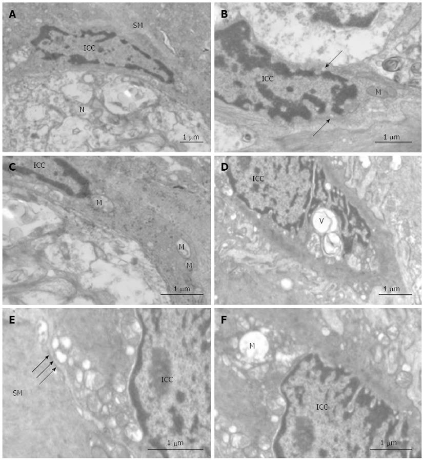
Figure 5 Ultrastructure of interstitial cells of Cajal.
A-C: Control. A: Interstitial cells of Cajal (ICC) with fusiform nuclear morphology show an elongated nucleus with scarce perinuclear cytoplasm, and are situated between the smooth muscle (SM) and the enteric nerve (N). The cytoplasmic processes surround the external contours of the enteric nerve; B: Caveolae are lining cytoplasmic membrane (arrows); C: The processes of some ICC have characteristically numerous mitochondria (M); D-F: Severe acute pancreatitis. D: Vacuoles (V) are present in the ICC; E: The density of desmosome-like junction between ICC and smooth muscle is lower (arrows); F: Vacuolated mitochondria are present in the processes of ICC.
- Citation: Shi LL, Liu MD, Chen M, Zou XP. Involvement of interstitial cells of Cajal in experimental severe acute pancreatitis in rats. World J Gastroenterol 2013; 19(14): 2179-2186
- URL: https://www.wjgnet.com/1007-9327/full/v19/i14/2179.htm
- DOI: https://dx.doi.org/10.3748/wjg.v19.i14.2179


