Copyright
©2013 Baishideng Publishing Group Co.
World J Gastroenterol. Apr 14, 2013; 19(14): 2144-2153
Published online Apr 14, 2013. doi: 10.3748/wjg.v19.i14.2144
Published online Apr 14, 2013. doi: 10.3748/wjg.v19.i14.2144
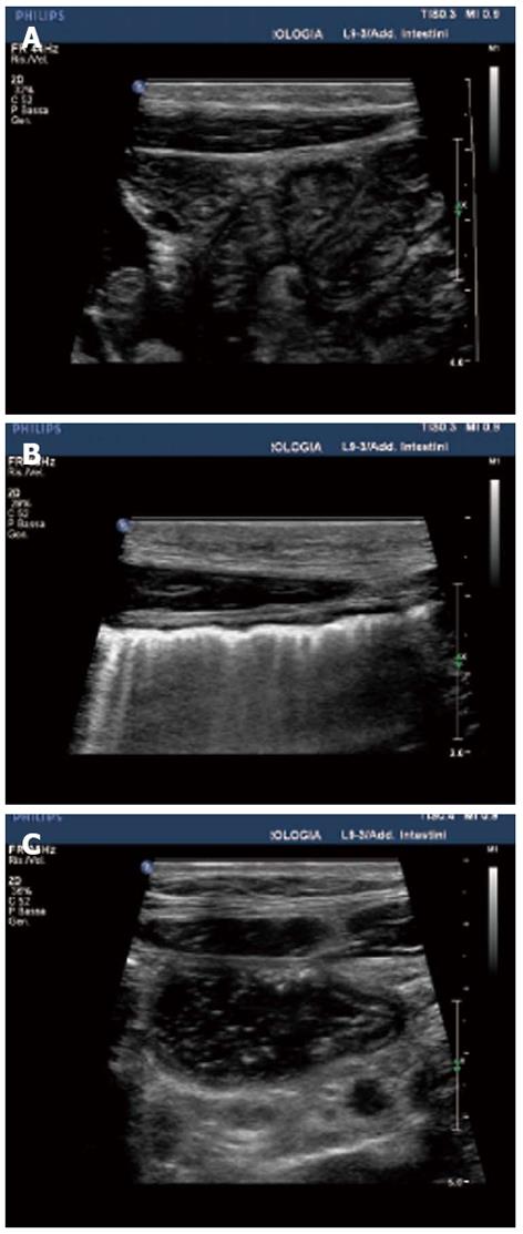
Figure 1 Sonographic appearance of normal bowel.
A: Mucus pattern: collapsed bowel containing only a highly reflective core of mucus with target appearance on a transverse section; B: Gas pattern: only the proximal side of the bowel wall is visible due to beam attenuation by gas; C: Fluid pattern: the bowel is filled with fluid and faeces with a tubular appearance on a longitudinal section.
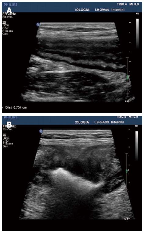
Figure 2 Wall thickening.
A: Inflammatory thickening: regular, with preserved wall stratification; B: Neoplastic thickening: irregular with “pseudokidney appearance”.
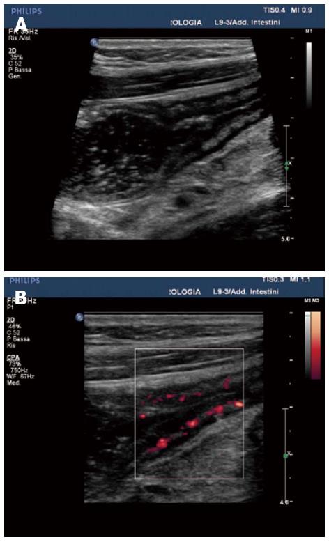
Figure 3 Stenosis in patients with Crohn’s disease.
A: B-mode aspect: narrow lumen with dilatation of the upstream segments; B: The presence of vascular signals on power Doppler indicates the inflammatory nature of stenosis.
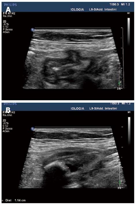
Figure 4 Diverticular disease.
A: Reflective outpouchings adjacent to the colonic wall; B: Acoustic shadowing outside the lumen indicating the presence of a coprolith.
- Citation: Roccarina D, Garcovich M, Ainora ME, Caracciolo G, Ponziani F, Gasbarrini A, Zocco MA. Diagnosis of bowel diseases: The role of imaging and ultrasonography. World J Gastroenterol 2013; 19(14): 2144-2153
- URL: https://www.wjgnet.com/1007-9327/full/v19/i14/2144.htm
- DOI: https://dx.doi.org/10.3748/wjg.v19.i14.2144


