Copyright
©2013 Baishideng Publishing Group Co.
World J Gastroenterol. Mar 14, 2013; 19(10): 1661-1664
Published online Mar 14, 2013. doi: 10.3748/wjg.v19.i10.1661
Published online Mar 14, 2013. doi: 10.3748/wjg.v19.i10.1661
Figure 1 Computed tomography showing the location of a potential short stricture (arrow) in the preterminal ileum with mild pre- and poststenotic dilatation.
This stricture was identified only after second reading of the computed tomography images and based upon the enteroscopic findings. A: Transverse plane; B: Coronal plane; C: Parasagittal plane.
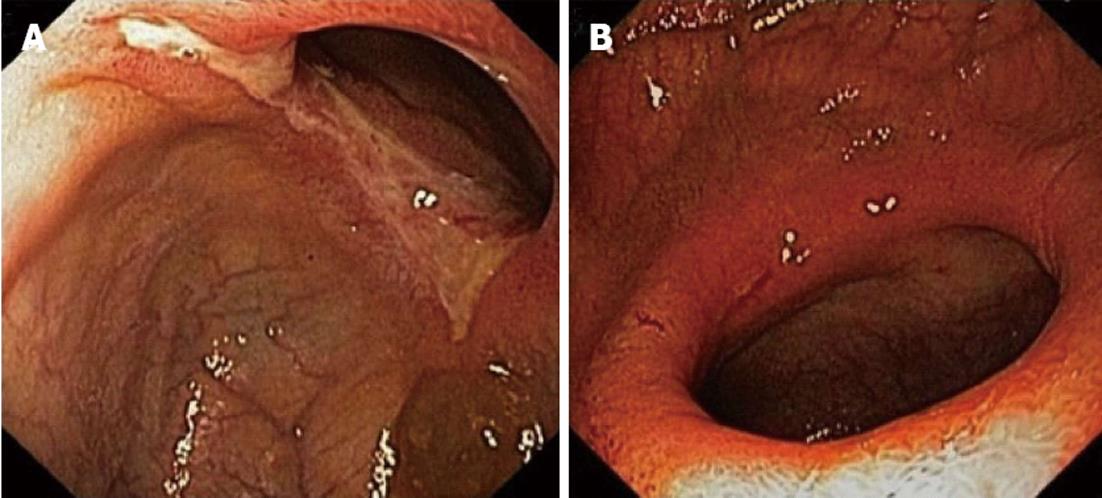
Figure 2 High definition endoscopic image of a circular ulcerative stenosis in the ileum, 50 cm proximal from the ileocecal valve.
After infliximab induction therapy, the ulceration almost completely disappeared and only a short hyperemic and less pronounced stenosis remained. A: Before infliximab; B: After infliximab.
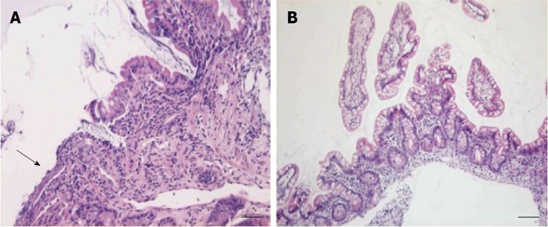
Figure 3 Ileal biopsy before and after infliximab treatment.
A: Superficial ulceration of the mucosa (arrow), with an acute inflammatory infiltrate in the lamina propria (HE stained paraffin section, bar: 100 μm); B: Ileal biopsy after treatment with infliximab, showing restoration of the villus architecture and only slight, non-specific inflammatory changes (bar: 200 μm).
- Citation: De Schepper H, Macken E, Van Marck V, Spinhoven M, Pelckmans P, Moreels T. Infliximab induces remission in cryptogenic multifocal ulcerous stenosing enteritis: First case. World J Gastroenterol 2013; 19(10): 1661-1664
- URL: https://www.wjgnet.com/1007-9327/full/v19/i10/1661.htm
- DOI: https://dx.doi.org/10.3748/wjg.v19.i10.1661


