Copyright
©2013 Baishideng Publishing Group Co.
World J Gastroenterol. Jan 7, 2013; 19(1): 108-114
Published online Jan 7, 2013. doi: 10.3748/wjg.v19.i1.108
Published online Jan 7, 2013. doi: 10.3748/wjg.v19.i1.108
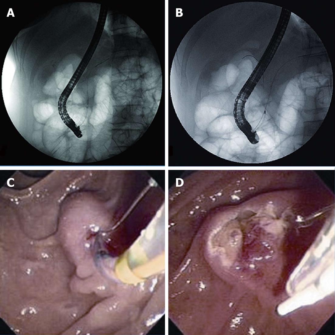
Figure 1 Radiologic images showing the use of double-guidewire technique and transpancreatic precut sphincterotomy.
A: Guidewire inserted and left in the pancreatic duct (PD) [double-guidewire technique (DGT)]; B: Common bile duct cannulation with a guidewire after previous insertion of a guidewire in the PD (DGT); C: Sphincterotomy performed with a cutting wire along the biliary direction at 11 o’clock [transpancreatic precut sphincterotomy (TPS)] with guidewire inserted and left in the PD (TPS); D: The bile duct orifice exposed to the left and below the pancreatic orifice (TPS). And then common bile duct cannulation after transpancreatic sphincterotomy.
Figure 2 Subject flow in the study.
CBD: Common bile duct; DGT: Double-guidewire technique; TPS: Transpancreatic sphincterotomy.
-
Citation: Yoo YW, Cha SW, Lee WC, Kim SH, Kim A, Cho YD. Double guidewire technique
vs transpancreatic precut sphincterotomy in difficult biliary cannulation. World J Gastroenterol 2013; 19(1): 108-114 - URL: https://www.wjgnet.com/1007-9327/full/v19/i1/108.htm
- DOI: https://dx.doi.org/10.3748/wjg.v19.i1.108


