Copyright
©2012 Baishideng Publishing Group Co.
World J Gastroenterol. Mar 7, 2012; 18(9): 971-978
Published online Mar 7, 2012. doi: 10.3748/wjg.v18.i9.971
Published online Mar 7, 2012. doi: 10.3748/wjg.v18.i9.971
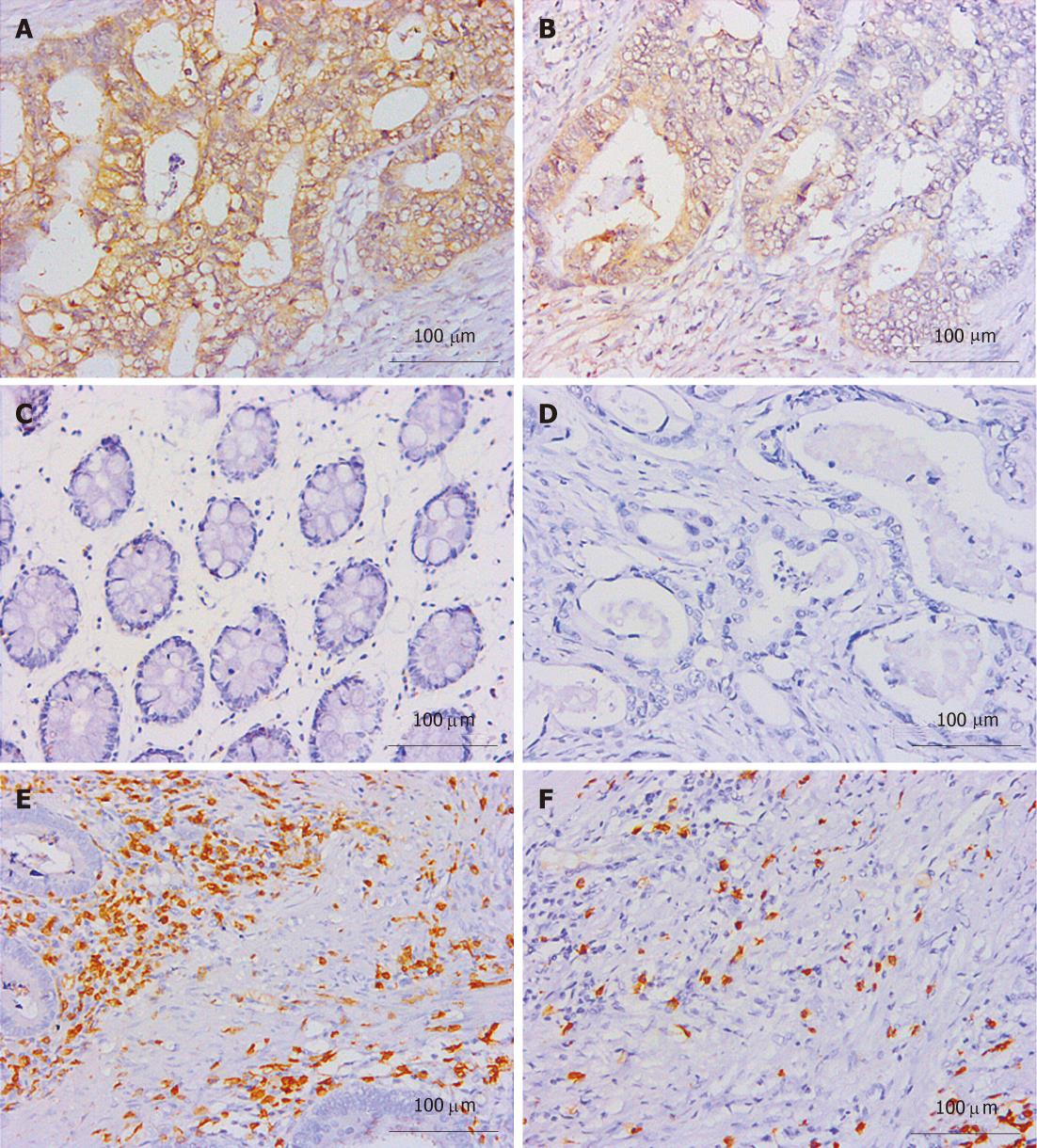
Figure 1 B7-H1 and CD3 immunostaining of colorectal carcinoma tissues.
A and B: B7-H1 immunostaining in colorectal carcinoma tissues (A: Magnification 400 ×; B: Magnification 200 ×); C: B7-H1 immunostaining in normal colorectal tissues; D: Negative control in colorectal carcinoma tissues; E and F: CD3 stained infiltrating T lymphocytesb (E: Tumor stroma; F: Tumor nest).
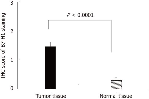
Figure 2 B7-H1 expression level in colorectal carcinoma tissues and adjacent normal tissues from 33 patients evaluated by immunohistochemisty.
IHC: Immunohistochemisty.
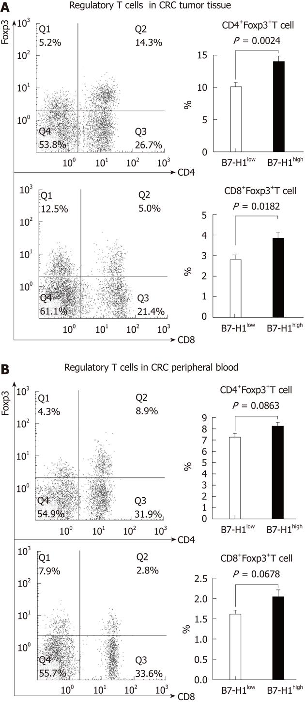
Figure 3 An elevated CD4+Foxp3+ and CD8+Foxp3+T cell amount observed in B7-H1high colorectal carcinoma tissues.
Mononuclear cells were harvested from fresh tumor tissues (A) and the peripheral blood (B) of the same colorectal carcinoma patient. The percentage of the Foxp3+ T cells was determined by fluorescence-activated cell sorting analysis. A: The population of CD4+Foxp3+ and CD8+Foxp3+T cells was increased remarkably in B7-H1high colorectal carcinoma (CRC) tissues compared with B7-H1low CRC tissues; B: In peripheral blood, there was no significant diversity of regulatory T cells between the B7-H1low and B7-H1high CRC patients.
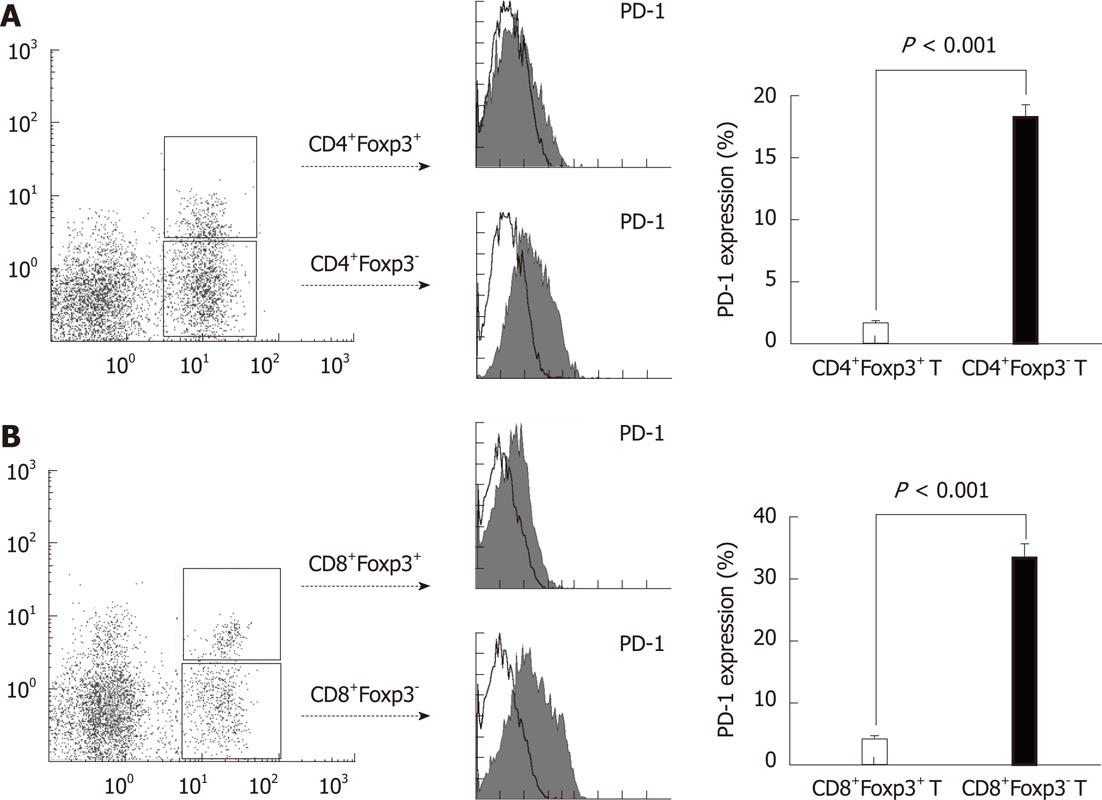
Figure 4 PD-1 expression on CD4+Foxp3+ or CD8+Foxp3+ regulatory T cells and conventional T cells in colorectal carcinoma tissues.
Mononuclear cells were harvested from fresh tumor tissues of the same colorectal carcinoma patient. The PD-1 expression was determined by fluorescence-activated cell sorting analysis, gated on CD4+Foxp3+/- or CD8+Foxp3+/- T cells. A: CD4+Foxp3+ regulatory T cells could hardly express PD-1 on the cell surface, while PD-1 expression level was significantly higher on the conventional CD4+T cells; B: CD8+Foxp3+ regulatory T cells almost failed to express PD-1, while PD-1 expression was significantly higher on the conventional CD8+T cells as well.
- Citation: Hua D, Sun J, Mao Y, Chen LJ, Wu YY, Zhang XG. B7-H1 expression is associated with expansion of regulatory T cells in colorectal carcinoma. World J Gastroenterol 2012; 18(9): 971-978
- URL: https://www.wjgnet.com/1007-9327/full/v18/i9/971.htm
- DOI: https://dx.doi.org/10.3748/wjg.v18.i9.971


