Copyright
©2012 Baishideng Publishing Group Co.
World J Gastroenterol. Mar 7, 2012; 18(9): 896-904
Published online Mar 7, 2012. doi: 10.3748/wjg.v18.i9.896
Published online Mar 7, 2012. doi: 10.3748/wjg.v18.i9.896
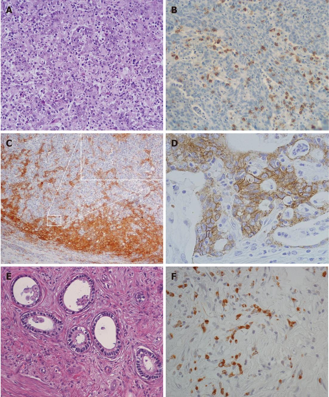
Figure 1 Histological and histochemical aspects of high lymphoid response cancers.
A: Lymphoepithelioid pattern of an HLR EBV+ tumor (hematoxylin-eosin, × 200); B: High intratumor T8 cells infiltration in an HLR EBV-/MSI- case (CD8 immunoperoxidase-hematoxylin, × 200); C: Peritumor dendritic cell rich demarcating band in an HLR MSI-H tumor (CD11c immunoperoxidase-hematoxylin, × 100); in the inset, focal enlargement of image C to show interaction of CD11c-reactive dendritic cells with unreactive neoplastic cells (× 400); D: HLA-DR reactivity of an HLR MSI-H case (immunoperoxidase-hematoxylin, × 400); E: Low-grade very-well-differentiated tubular carcinoma (hematoxylin-eosin, × 200); F: Low-grade desmoplastic tumor with spindle neoplastic cells interspersed among fibroblast rich stroma (CAR5, immunoperoxidase-hematoxylin, × 200).
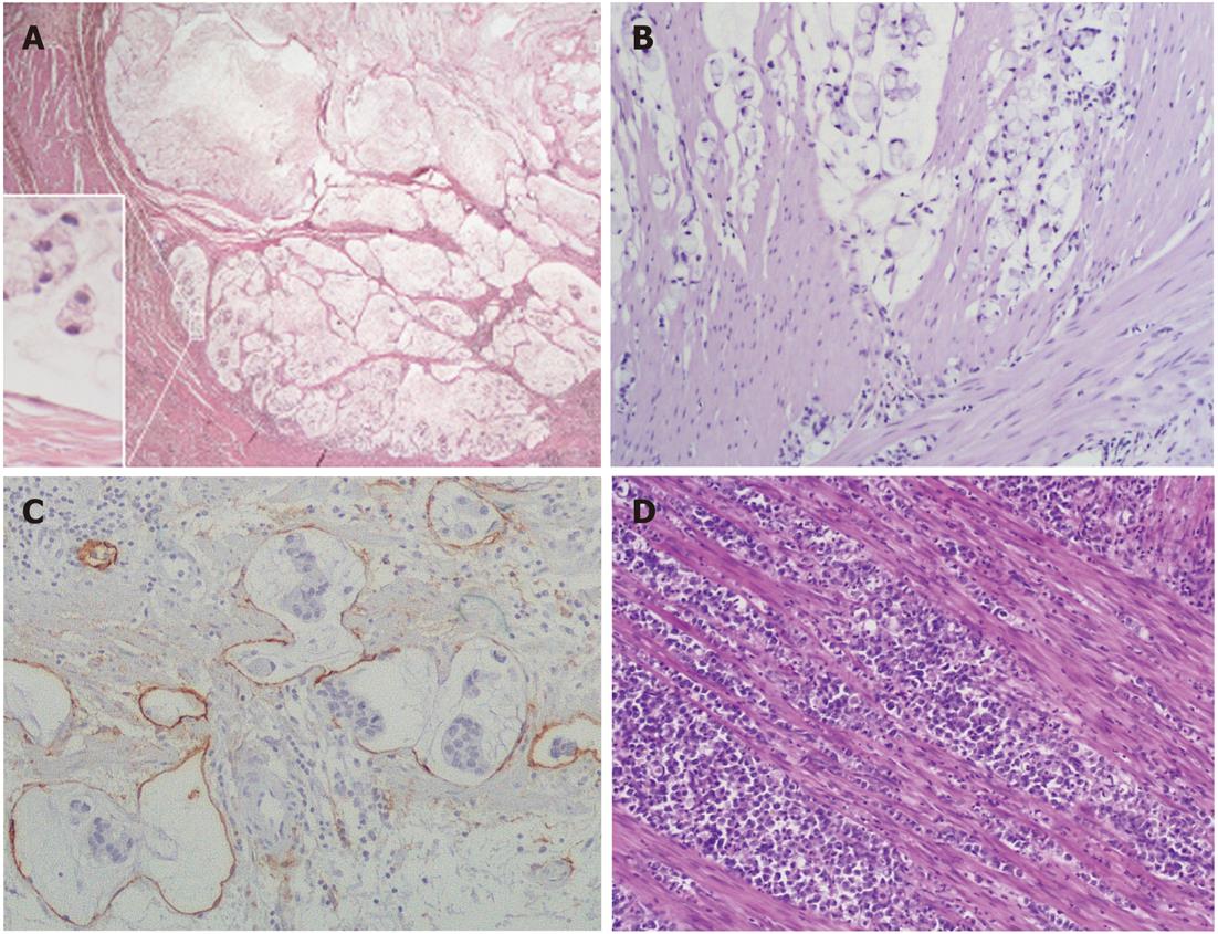
Figure 2 Muconodular, well-differentiated tubular, diffuse desmoplastic and high lymphoid response.
A: Low-grade muconodular cancer with expansile growth (hematoxylin-eosin, × 20); in the inset, note tumor cells floating inside mucin, free of contact with stroma (× 400); B: intermediate-grade, locally infiltrative mucinous cancer (hematoxylin-eosin, × 200); C: massive lymphoinvasion of a high-grade mucinous cancer (podoplanin immunoperoxidase-hematoxylin, × 200); D: Diffuse anaplastic cancer invading the muscularis propria (hematoxylin-eosin, × 200), in the inset the enlargement shows cellular pleomorphism (× 400) .
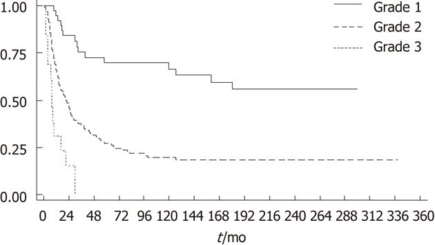
Figure 3 Kaplan-Meier survival estimate of 200 gastric cancers according to histotype-based grade.
- Citation: Chiaravalli AM, Klersy C, Vanoli A, Ferretti A, Capella C, Solcia E. Histotype-based prognostic classification of gastric cancer. World J Gastroenterol 2012; 18(9): 896-904
- URL: https://www.wjgnet.com/1007-9327/full/v18/i9/896.htm
- DOI: https://dx.doi.org/10.3748/wjg.v18.i9.896


