Copyright
©2012 Baishideng Publishing Group Co.
World J Gastroenterol. Feb 28, 2012; 18(8): 840-846
Published online Feb 28, 2012. doi: 10.3748/wjg.v18.i8.840
Published online Feb 28, 2012. doi: 10.3748/wjg.v18.i8.840
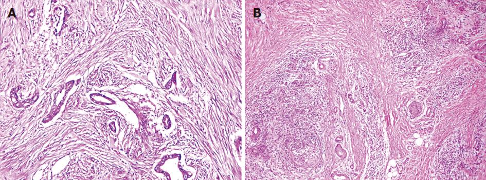
Figure 1 Fibrotic focus in pancreatic ductal adenocarcinoma.
A: In the fibrotic focus, fibroblasts and collagen fibers are radially aligned around the tumor cells (× 200); B: A pattern different from that in the surrounding pancreatic ductal adenocarcinoma stroma where the fibroblasts and collagen fibers are arranged in an orderly fashion (× 100).
Figure 2 Fibroblast activation protein expression in pancreatic ductal adenocarcinoma.
A: Fibroblast activation protein (FAP) expression in carcinoma cells is located mainly on the cell membrane and in the cytoplasm (× 200); B: FAP-positive fibroblasts are mainly located adjacent to carcinoma tissue (× 200); C: FAP is expressed in carcinoma cells and in fibroblasts adjacent to carcinoma tissue (× 100); D: The adjacent noncancerous pancreatic ducts show no positive staining for FAP and fibroblasts away from the tumor tissues rarely express FAP (× 200).
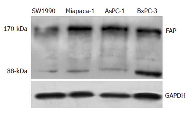
Figure 3 Western blotting analysis of fibroblast activation protein in pancreatic cancer cell lines.
All four cell lines express fibroblast activation protein (FAP) with protein bands corresponding to the proteolytically active 170-kDa seprase dimer and its 88-kDa seprase subunit protein. GAPDH: Glyceraldehyde-3-phosphate dehydrogenase.
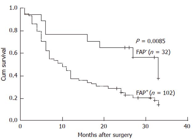
Figure 4 Kaplan-Meier survival curves of pancreatic ductal adenocarcinoma patients.
The prognosis was significantly worse in the fibroblast activation protein (FAP)-positive group compared with the FAP-negative group (P = 0.0085).
- Citation: Shi M, Yu DH, Chen Y, Zhao CY, Zhang J, Liu QH, Ni CR, Zhu MH. Expression of fibroblast activation protein in human pancreatic adenocarcinoma and its clinicopathological significance. World J Gastroenterol 2012; 18(8): 840-846
- URL: https://www.wjgnet.com/1007-9327/full/v18/i8/840.htm
- DOI: https://dx.doi.org/10.3748/wjg.v18.i8.840


