Copyright
©2012 Baishideng Publishing Group Co.
World J Gastroenterol. Feb 7, 2012; 18(5): 435-444
Published online Feb 7, 2012. doi: 10.3748/wjg.v18.i5.435
Published online Feb 7, 2012. doi: 10.3748/wjg.v18.i5.435
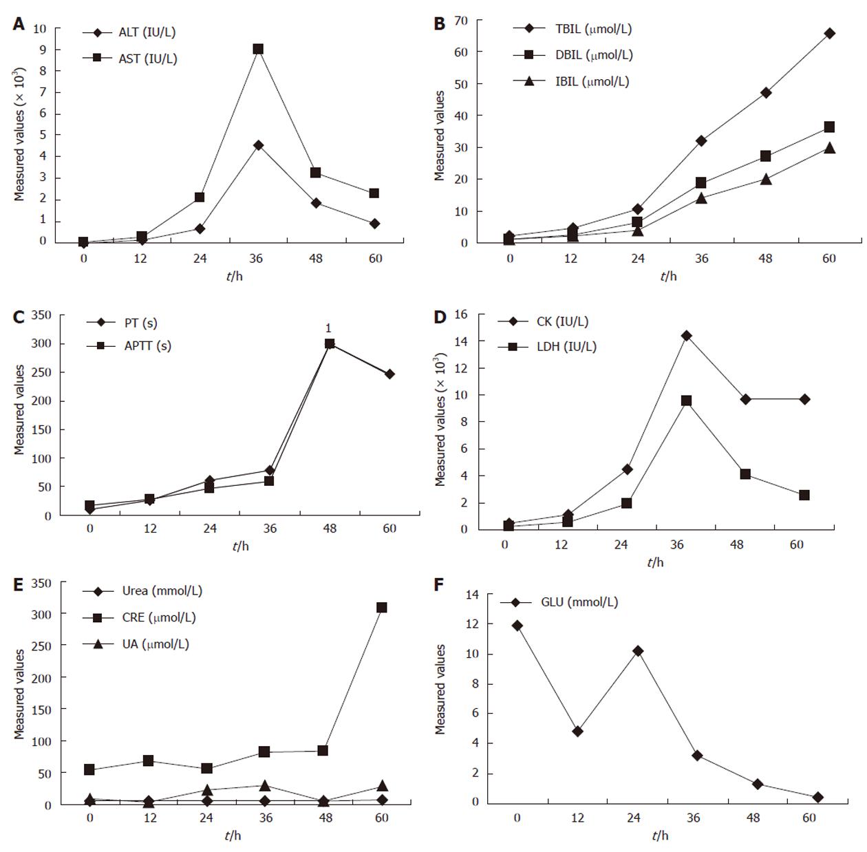
Figure 1 Blood biochemical parameters of the Macaca mulatta model of fulminant hepatic failure.
The abscissa and ordinate represent measured time and value, respectively. ALT: Alanine aminotransferase; AST: Aspartate aminotransferase; TBIL: Total bilirubin; DBIL: Direct bilirubin; IBIL: Indirect bilirubin; PT: Prothrombin time; APTT: Activated partial thromboplastin time; CK: Creatine kinase; LDH: Lactate dehydrogenase; CRE: Creatinine; UA: Uric acid; GLU: Glucose; FHF: Fulminant hepatic failure. 1Represents values > 300 s, which exceeded the range of the equipment, so we did not obtain an exact measurement.
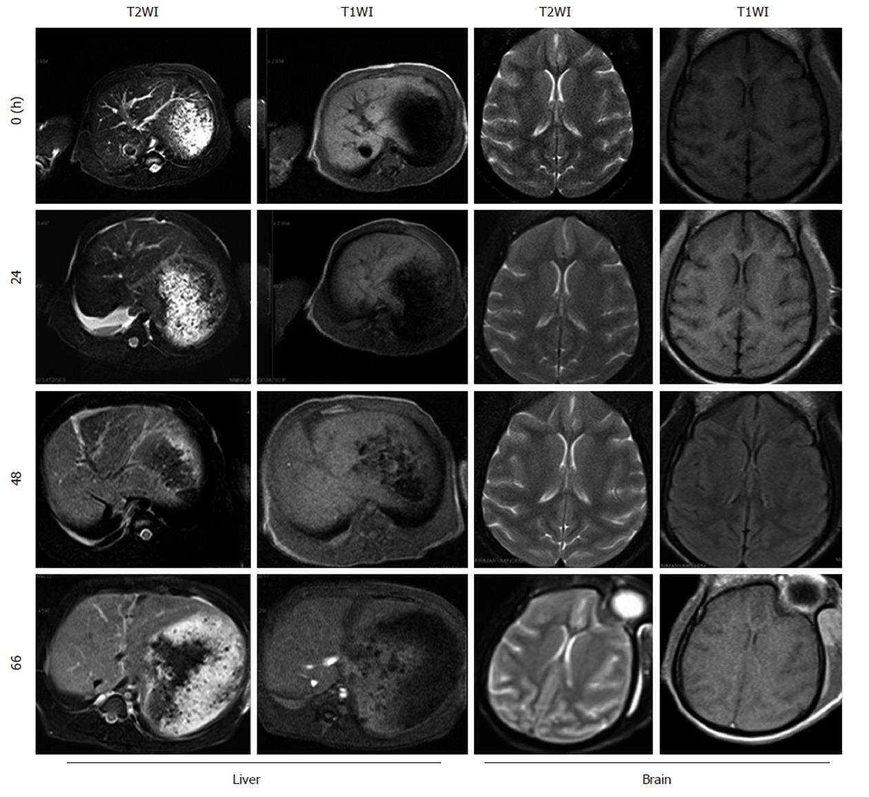
Figure 2 Magnetic resonance imaging of the liver and brain of Macaca mulatta model at different times.
T2WI: T2-weighted imaging; T1WI: T1-weighted imaging.
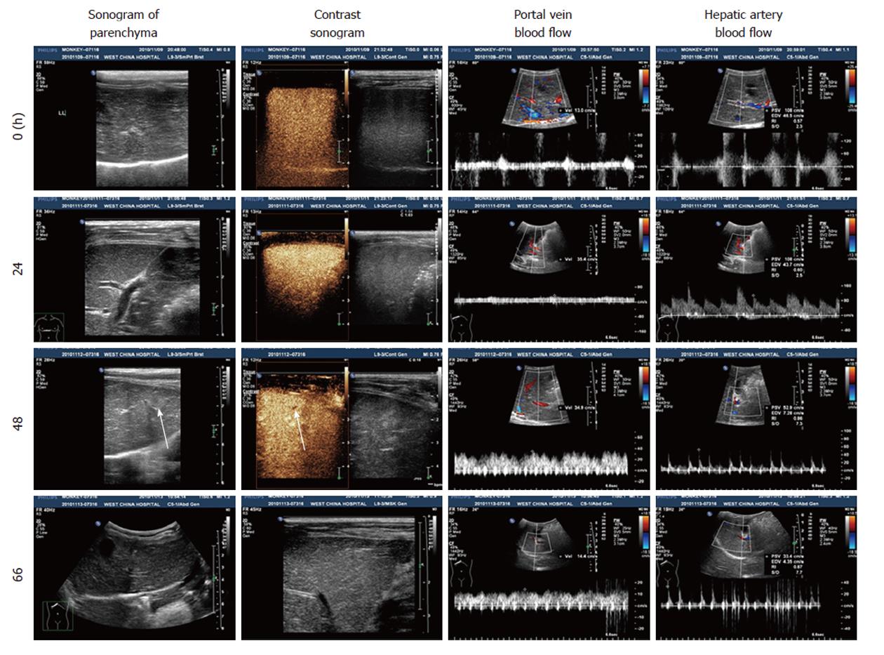
Figure 3 Liver sonogram of the Macaca mulatta model at different times.
Arrows show a slightly enhanced echo and lower perfusion of SonoVue at 48 h.
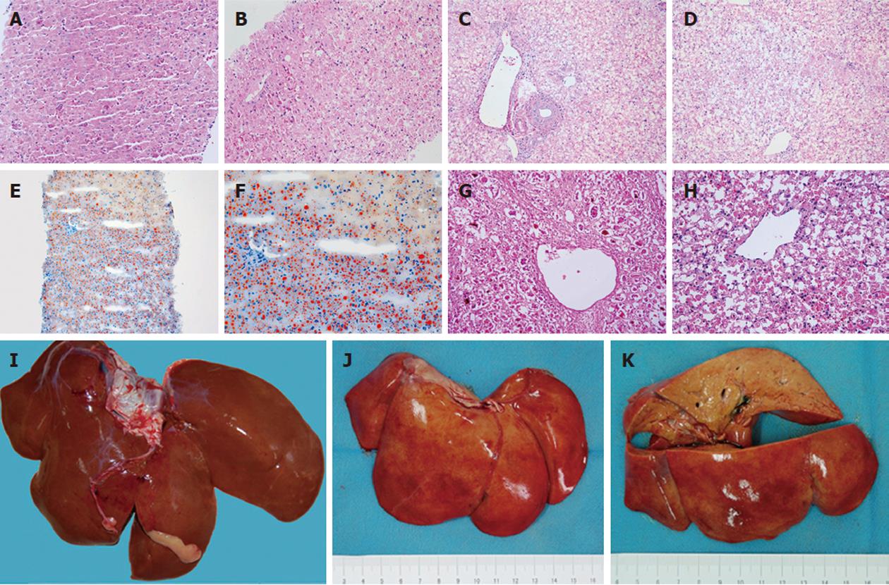
Figure 4 Histopathological changes of the liver.
A: Tissue from needle biopsy at 12 h. Changes in organizational structure and cell morphology were not obvious [hematoxylin and eosin (H and E) stain, × 200]; B: Tissue from needle biopsy at 36 h. Vacuoles appeared in the hepatocellular cytoplasm (H and E stain, × 200); C, D: Tissue from necropsy after death at 70 h. Hepatocellular necrosis was distributed in the portal area (C) and central area (D) (H and E stain, × 200). Massive necrosis caused almost all of the hepatocytes to be lost, and only the support structure remained; E: Frozen tissue from needle biopsy at 36 h. The extensive reddish-orange color suggested serious fatty degeneration (oil red O stain, × 100); F: Frozen tissue from needle biopsy at 36 h. In the borderline between necrosis and steatosis, the reddish-orange color was obvious (oil red O stain, × 200); G, H: Comparison of pathological changes of fulminant hepatic failure in humans induced by viral hepatitis (G) and a Macaca mulatta model induced by α-amanitin and lipopolysaccharide (H) (H and E stain, × 200); I: Normal liver of Macaca mulatta for comparison; J: The surface view of the experimental liver on necropsy appeared red and yellow; K: The cut surface view of the experimental liver was uniformly yellow.
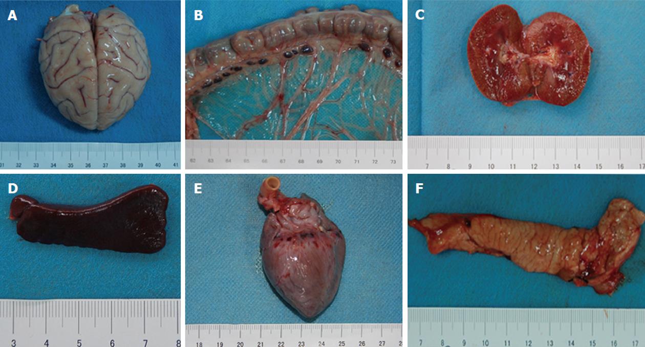
Figure 5 Other main organs on necropsy.
A: The brain with cerebral edema; B: Mesenteric lymph nodes enlarged and beaded; Hyperemia of the kidney (C), spleen (D), heart (E), and pancreas (F) without obvious gross lesions.
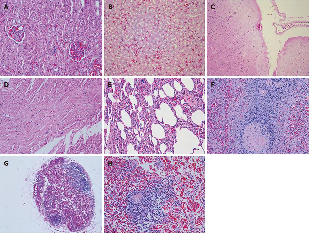
Figure 6 Histopathological changes of other organs.
A: Ectasia and hyperemia of capillaries in the renal glomerulus [hematoxylin and eosin (H and E) stain, × 200]; B: Widespread hyperemia of capillaries in the renal stroma and cellular swelling in the tubular epithelial cells (H and E stain, × 100); C: Weak staining in the superficial layer of the brain suggest cerebral edema (H and E stain, × 100); D: Wave-like changes in the myocardium (H and E stain, × 200); E: Hyperemia of capillaries in the stroma and hyaline changes in the arterioles of the lung (H and E stain, × 200); F: Hyaline changes in the central artery of the spleen (H and E stain, × 200); G: Widespread reduction in lymphocytes and hemorrhage in the mesenteric lymph nodes (H and E stain, × 50); H: Hemorrhage and histiocyte proliferation in the mesenteric lymph nodes. Hyaline change appeared in the vessel wall of lymphoid follicles (H and E stain, × 200).
-
Citation: Zhou P, Xia J, Guo G, Huang ZX, Lu Q, Li L, Li HX, Shi YJ, Bu H. A
Macaca mulatta model of fulminant hepatic failure. World J Gastroenterol 2012; 18(5): 435-444 - URL: https://www.wjgnet.com/1007-9327/full/v18/i5/435.htm
- DOI: https://dx.doi.org/10.3748/wjg.v18.i5.435


