Copyright
©2012 Baishideng Publishing Group Co.
World J Gastroenterol. Dec 14, 2012; 18(46): 6836-6842
Published online Dec 14, 2012. doi: 10.3748/wjg.v18.i46.6836
Published online Dec 14, 2012. doi: 10.3748/wjg.v18.i46.6836
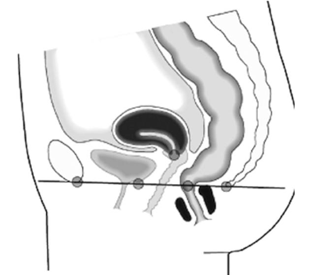
Figure 1 The pubococcygeal line (black) according to Yang et al[3] from the most inferior part of the pubic symphysis to the last coccygeal joint; furthermore, gray dots show (from left to right) bladder base, uterocervical junction, and anorectal junction.
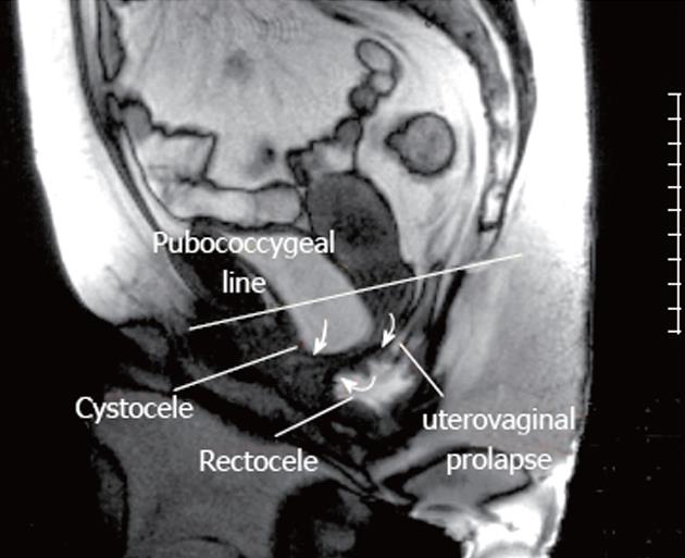
Figure 2 Dynamic magnetic resonance imaging of a 36-year-old healthy female volunteer.
At the end of defection, a rectocele, cystocele and uterovaginal prolapse were visualized.
- Citation: Schreyer AG, Paetzel C, Fürst A, Dendl LM, Hutzel E, Müller-Wille R, Wiggermann P, Schleder S, Stroszczynski C, Hoffstetter P. Dynamic magnetic resonance defecography in 10 asymptomatic volunteers. World J Gastroenterol 2012; 18(46): 6836-6842
- URL: https://www.wjgnet.com/1007-9327/full/v18/i46/6836.htm
- DOI: https://dx.doi.org/10.3748/wjg.v18.i46.6836


