Copyright
©2012 Baishideng Publishing Group Co.
World J Gastroenterol. Dec 14, 2012; 18(46): 6701-6708
Published online Dec 14, 2012. doi: 10.3748/wjg.v18.i46.6701
Published online Dec 14, 2012. doi: 10.3748/wjg.v18.i46.6701
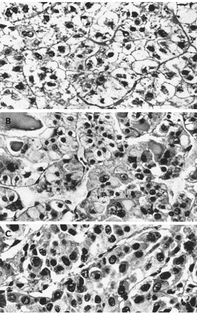
Figure 1 Light micrographs of portions from human hepatocellular neoplasms with and without glycogenosis.
A: Clear-cell hepatocellular adenoma consisting predominantly of glycogenotic cells. In some cells (arrows) there is a reduction of glycogen and focal increase in cytoplasmic basophilia; B: Highly differentiated hepatocellular carcinoma (HCC) composed of a mixed population of clear (glycogenotic) cells, acidophilic cells (ground-glass hepatocytes, arrows), and some glycogen-poor, basophilic cells; C: Poorly differentiated, glycogen-free, basophilic HCC. All: Hematoxylin and eosin stain, x 460, from Bannasch et al[6].
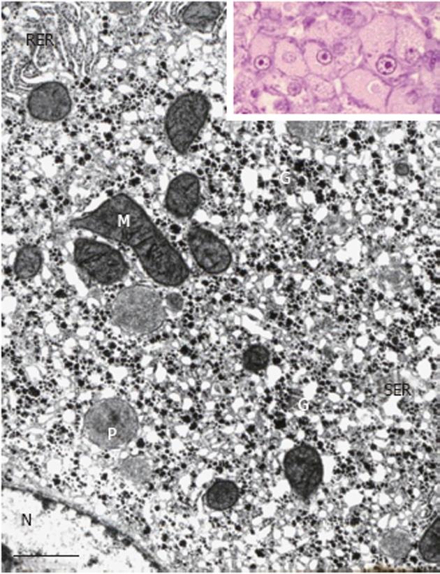
Figure 2 Portion of a glycogenotic acidophilic hepatocyte (corresponding to glycogen-ground-glass hepatocyte) induced in rat liver by N-nitrosomorpholine.
Note abundant α- and β-glycogen particles (G) in close spatial relationship with large network complexes of proliferated smooth endoplasmic reticulum (SER). Rough endoplasmic reticulum (RER), mitochondria (M), peroxisomes (P), and nucleus (N). Inset: Group of acidophilic hepatocytes as seen under the light microscope. Transmission electron microscopy, lead citrate. Bar: 1 μm.
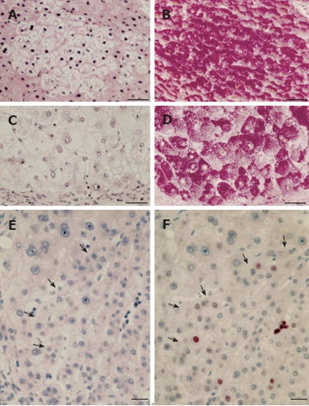
Figure 3 Human focal (A, B) and nodular (C, D) hepatic glycogenosis, and more advanced mixed cell populations (E, F) with intrafocal small cell change.
A, B: Hepatic vein. Perivenular glycogenolytic (clear cell) focus in an hepatocellular carcinoma (HCC)-bearing liver with hepatitis B virus (HBV)-associated cirrhosis, demonstrated in serial sections with hematoxylin and eosin (A) and periodic acid schiff (PAS) (B)-reaction counterstained with orange G and iron hematoxilin (Tri-PAS). Bar: 100 μm; C, D: Glycogenotic (clear cell) nodule in an HCC-free liver with HBV-associated cirrhosis demonstrated in serial sections with hematoxylin-eosin (C) and Tri-PAS (D). Bar: 100 μm; E, F: Hematoxylin and eosin (E), proliferating cell nuclear antigen (F) immunostaining. Increased cell proliferation (arrows) in a mixed cell focus with less pronounced glycogen storage (not shown) and with intrafocal small cell change in liver with cryptogenic cirrhosis, demonstrated in serial sections. Bar: 50 μm, from Su et al[68].
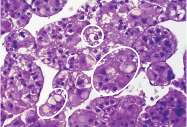
Figure 4 Portion of a human trabecular hepatocellular carcinoma with mixed populations, including clear (glycogenotic) cells (small upper white circle), acidophilic (glycogenotic) ground-glass cells (somewhat larger white circle at the bottom, left), transitions from ground-glass hepatocytes into basophilic cell populations (largest white circle in the middle, right), and basophilic cell populations in the middle and at the bottom right (not marked by circles).
Hematoxylin and eosin.
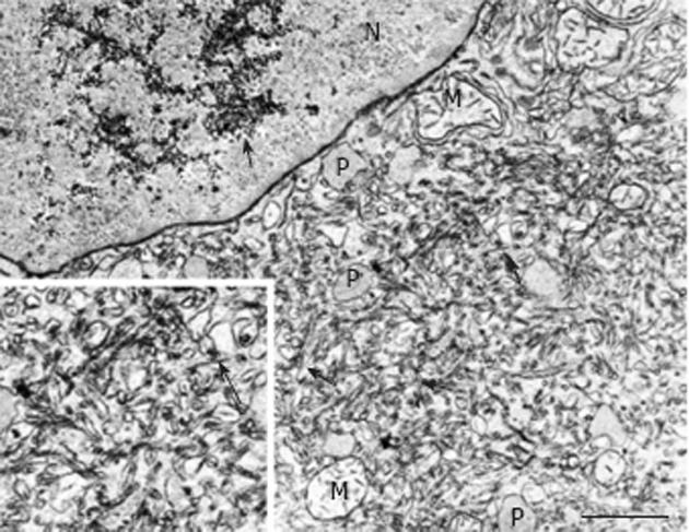
Figure 5 Portion of a ground-glass hepatocyte from a human liver with hepatitis B virus-associated cirrhosis, showing an accumulation of glycogen granules (long arrow) in the nucleus (N) and abundant smooth endoplasmic reticulum containing filamentous hepatitis B surface antigen (short arrows).
M: Mitochondria: P: Peroxisome. Transmission electron microscopy, lead citrate. Bar: 1 μm.
- Citation: Bannasch P. Glycogenotic hepatocellular carcinoma with glycogen-ground-glass hepatocytes: A heuristically highly relevant phenotype. World J Gastroenterol 2012; 18(46): 6701-6708
- URL: https://www.wjgnet.com/1007-9327/full/v18/i46/6701.htm
- DOI: https://dx.doi.org/10.3748/wjg.v18.i46.6701


