Copyright
©2012 Baishideng Publishing Group Co.
World J Gastroenterol. Dec 7, 2012; 18(45): 6682-6685
Published online Dec 7, 2012. doi: 10.3748/wjg.v18.i45.6682
Published online Dec 7, 2012. doi: 10.3748/wjg.v18.i45.6682
Figure 1 A computed tomography scan revealed a low-density heterogeneous mass of 81 mm × 68 mm in size in the tail of the pancreas (arrow) that invaded the greater curvature of the stomach and the spleen.
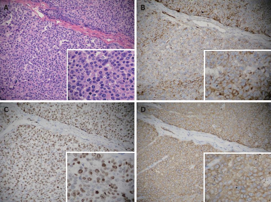
Figure 2 Pathology of the pancreas.
A: Spindle-shaped cells with scanty cytoplasm and hyperchromatic nuclei (hematoxylin and eosin staining); B: Positively stained for chlorhexidine A; C: Approximately 80% of the tumor cells were positively stained for Ki67; D: Tumor cells were positively stained for synaptophysin (B, C and D: EnVision) (original magnification: ×200 and ×400).
Figure 3 Positron emission tomography-computed tomography revealed high fludeoxyglucose uptake in the left leg and the relapse of carcinoma in both hila of the lungs.
Figure 4 Pathology of the leg.
A: Tumor cells displayed infiltrative growth in the striated muscle of the left leg [hematoxylin and eosin (HE) staining, original magnification: ×100]; B: Tumor cells were homogeneous (HE staining, original magnification: ×400); C: Tumor cells were positively stained for synaptophysin (EnVision, original magnification: ×400).
- Citation: Chen J, Zheng Q, Yang Z, Huang XY, Yuan Z, Tang J. Neuroendocrine carcinoma of the pancreas with soft tissue metastasis. World J Gastroenterol 2012; 18(45): 6682-6685
- URL: https://www.wjgnet.com/1007-9327/full/v18/i45/6682.htm
- DOI: https://dx.doi.org/10.3748/wjg.v18.i45.6682


