Copyright
©2012 Baishideng Publishing Group Co.
World J Gastroenterol. Dec 7, 2012; 18(45): 6577-6586
Published online Dec 7, 2012. doi: 10.3748/wjg.v18.i45.6577
Published online Dec 7, 2012. doi: 10.3748/wjg.v18.i45.6577
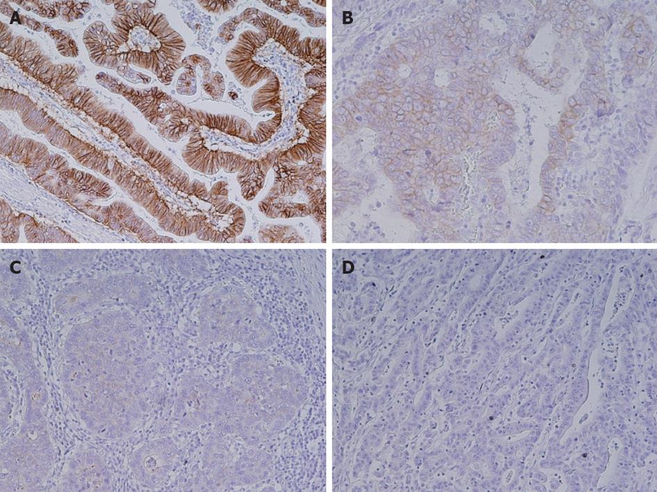
Figure 1 Immunohistochemical analysis of human epidermal growth factor receptor 2 in gastric cancer tissues.
A: Human epidermal growth factor receptor 2 (HER2) 3+; B: HER2 2+; C: HER2 1+; D: HER2 0. Original magnification, ×200.
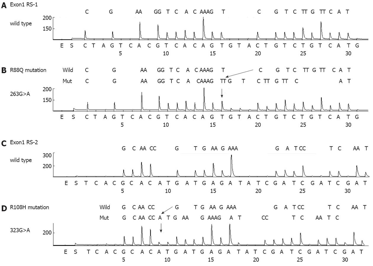
Figure 2 Phosphatidylinositol 3-kinase, catalytic, alpha polypeptide mutations detected by pyrosequencing in gastric cancer tissues.
A: Exon1 RS1 wild type; B: 263G>A (R88Q) mutation; C: Exon1 RS2 wild type; D: 323G>A (R108H) mutation.
Figure 3 In situ hybridization analysis of Epstein-Barr virus-encoded small RNA-1 and human epidermal growth factor receptor 2 immunohistochemical expression in gastric cancer tissues.
A: Gastric adenocarcinoma positive for Epstein-Barr virus-encoded small RNA-1 (EBER-1); B: Gastric adenocarcinoma negative for EBER-1; C: Immunohistochemical analysis of human epidermal growth factor receptor 2 (HER2) in an Epstein-Barr virus-positive and HER2-positive case. Original magnification, ×200.
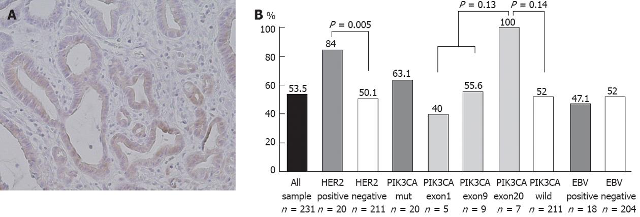
Figure 4 Immunohistochemical analysis and assessment of phospho Akt positivity based on molecular alterations in gastric cancer tissues.
A: Gastric adenocarcinoma showing phospho Akt (pAkt) positivity. Original magnification, ×200; B: pAkt expression significantly correlates with human epidermal growth factor receptor 2 (HER2) overexpression (P < 0.01) but not with phosphatidylinositol 3-kinase, catalytic, alpha polypeptide (PIK3CA) mutations (P = 0.37) or Epstein-Barr virus (EBV) infection (P = 0.69).
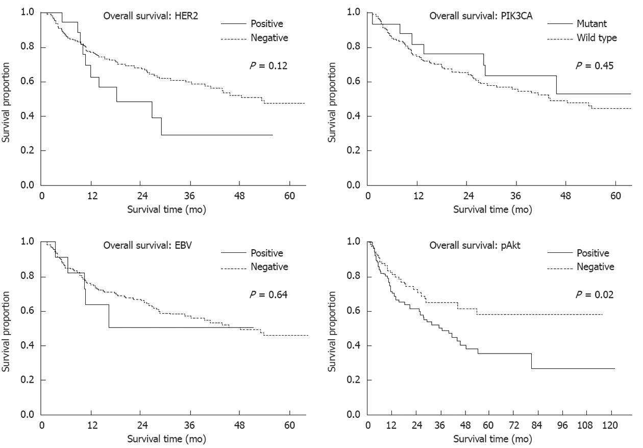
Figure 5 Survival analysis of gastric cancer patients.
Three year survival of human epidermal growth factor receptor 2 (HER2)-positive vs HER2-negative, 29.1 mo vs 59.4 mo; Phosphatidylinositol 3-kinase, catalytic, alpha polypeptide (PIK3CA) mutation vs wild type, 63.7 mo vs 56.3 mo; Epstein-Barr virus (EBV)-positive vs EBV-negative, 51.3 mo vs 57.6 mo; And phospho Akt (pAkt)-positive vs pAkt-negative, 50.7 mo vs 64.8 mo. Five year survival of pAkt-positive vs pAkt-negative cases, 35.5 mo vs 58.1 mo.
- Citation: Sukawa Y, Yamamoto H, Nosho K, Kunimoto H, Suzuki H, Adachi Y, Nakazawa M, Nobuoka T, Kawayama M, Mikami M, Matsuno T, Hasegawa T, Hirata K, Imai K, Shinomura Y. Alterations in the human epidermal growth factor receptor 2-phosphatidylinositol 3-kinase-v-Akt pathway in gastric cancer. World J Gastroenterol 2012; 18(45): 6577-6586
- URL: https://www.wjgnet.com/1007-9327/full/v18/i45/6577.htm
- DOI: https://dx.doi.org/10.3748/wjg.v18.i45.6577


