Copyright
©2012 Baishideng Publishing Group Co.
World J Gastroenterol. Nov 28, 2012; 18(44): 6515-6520
Published online Nov 28, 2012. doi: 10.3748/wjg.v18.i44.6515
Published online Nov 28, 2012. doi: 10.3748/wjg.v18.i44.6515
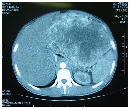
Figure 1 Abdominal computed tomography demonstrating a giant solitary tumor of 15.
8 cm × 21.0 cm in abdominal cavity.
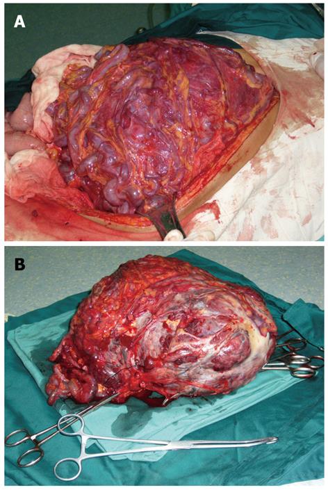
Figure 2 Giant tumor.
A: A giant tumor originating from greater omentum; B: A giant tumor originating from resected specimen.
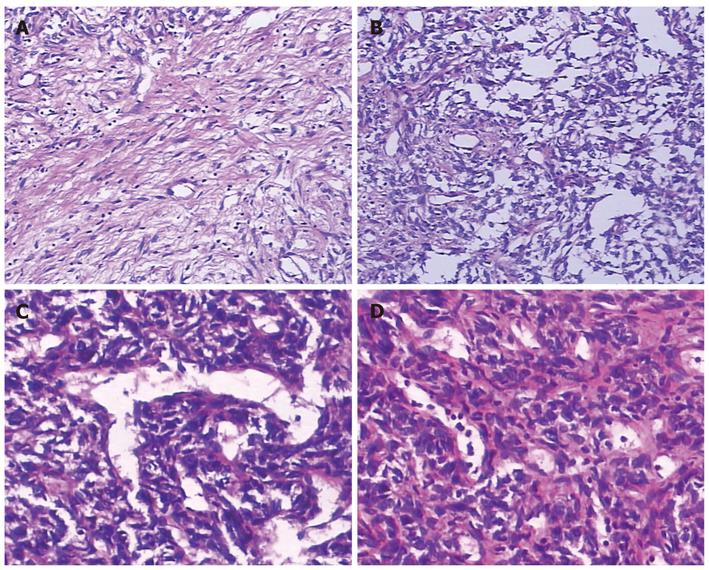
Figure 3 Hematoxylin and eosin stained sections.
A: Collagen deposition, 10 cm × 10 cm; B: Abundant spindle cells, 10 cm × 10 cm; C: Branching vessel, 10 cm × 20 cm; D: Nuclear atypia, 10 cm × 20 cm.
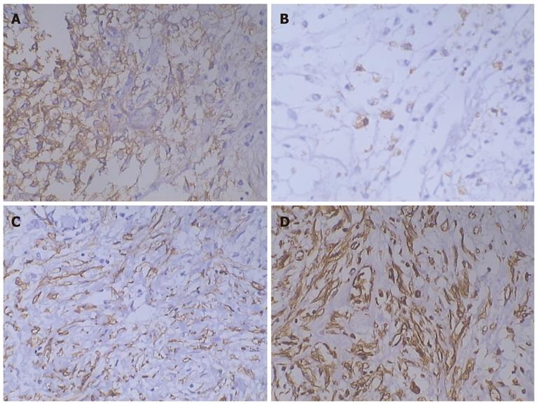
Figure 4 Immunohistochemical test.
Immunohistochemical test showing the tumor was positive for CD34 (A), bcl-2 (B), α-smooth muscle actin (C), and vimentin (D) (10 cm × 20 cm).
- Citation: Zong L, Chen P, Wang GY, Zhu QS. Giant solitary fibrous tumor arising from greater omentum. World J Gastroenterol 2012; 18(44): 6515-6520
- URL: https://www.wjgnet.com/1007-9327/full/v18/i44/6515.htm
- DOI: https://dx.doi.org/10.3748/wjg.v18.i44.6515


