Copyright
©2012 Baishideng Publishing Group Co.
World J Gastroenterol. Nov 28, 2012; 18(44): 6504-6509
Published online Nov 28, 2012. doi: 10.3748/wjg.v18.i44.6504
Published online Nov 28, 2012. doi: 10.3748/wjg.v18.i44.6504
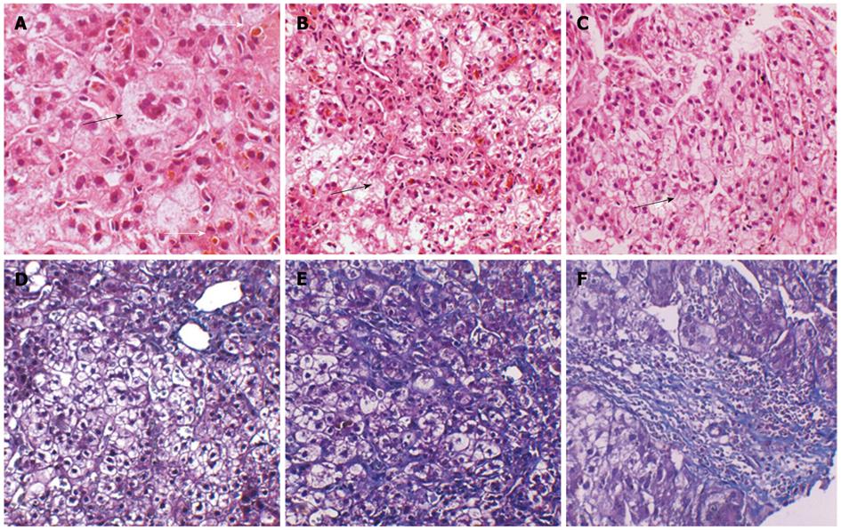
Figure 1 Histological features of samples taken at age 10 and current liver biopsy specimens (age 23 years).
A: Multinucleated giant cells (black arrow) and ductal cholestasis (white arrow) in the previously sampled liver tissue (image, × 400); B: Ductal cholestasis (white arrow) and ballooning degeneration of the hepatocytes (black arrow) in the previously sampled liver tissue (image, × 200); C: Ductal cholestasis and hepatocytes ballooning degeneration (black arrow) in the recently sampled liver tissue (image, × 200); D: Mild portal and lobular fibrosis in the previously sampled liver tissue (image, × 200); E: Moderate lobular fibrosis in the recently sampled liver tissue (image, × 200); F: Moderate portal fibrosis in the recently sampled liver tissue (image, × 200).
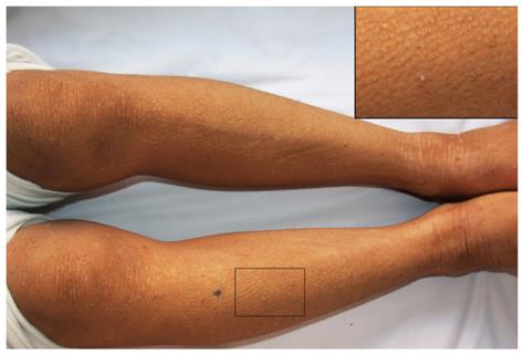
Figure 2 Skin.
The skin was rough and thickened.
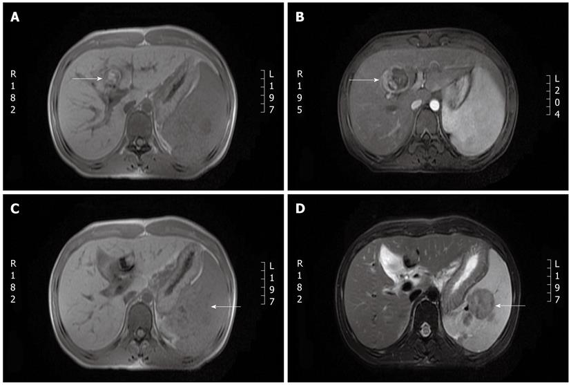
Figure 3 Liver magnetic resonance imaging.
A: A 2.3 cm × 2.56 cm lesion was observed in segment 4 of the liver (T1-weighted). No obvious dilation of the intra or extrahepatic bile duct was observed; B: Following contrast magnetic resonance imaging (MRI), the capsule shows linear enhancement without enhancement of the lesion itself. Splenomegaly is observed, and several oval-shaped nodules in the spleen show slight enhancement; C: The oval-shaped nodules appear as low-intensity signals on a T1-weighted MRI image; D: The oval-shaped nodules in the spleen appear as a lower-intensity signal on T2-weighted MRI images than on T1-weighted images.
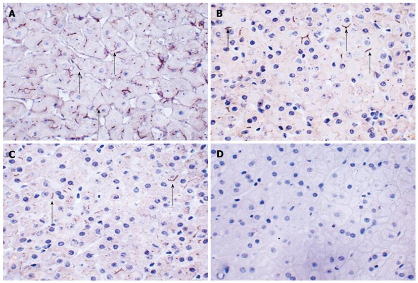
Figure 4 Expression pattern of the canalicular transporter bile salt export pump (rabbit anti-bile salt export pump polyclonal antibody, all images, × 400).
A: Normal bile salt export pump (BSEP) staining in a control liver without cholestasis. Anti-BSEP antibody stains an orderly canalicular network; B: BSEP staining in liver tissue collected from the patient 13 years ago. BSEP is present only focally (arrows) and is both faint and patchy; C: BSEP staining in the recent sample of liver tissue BSEP expression is again faint and patchy; D: Canaliculi in negative control liver stained with phosphate-buffered saline.
-
Citation: Deng BC, Lv S, Cui W, Zhao R, Lu X, Wu J, Liu P. Novel
ATP8B1 mutation in an adult male with progressive familial intrahepatic cholestasis. World J Gastroenterol 2012; 18(44): 6504-6509 - URL: https://www.wjgnet.com/1007-9327/full/v18/i44/6504.htm
- DOI: https://dx.doi.org/10.3748/wjg.v18.i44.6504


