Copyright
©2012 Baishideng Publishing Group Co.
World J Gastroenterol. Nov 21, 2012; 18(43): 6197-6205
Published online Nov 21, 2012. doi: 10.3748/wjg.v18.i43.6197
Published online Nov 21, 2012. doi: 10.3748/wjg.v18.i43.6197
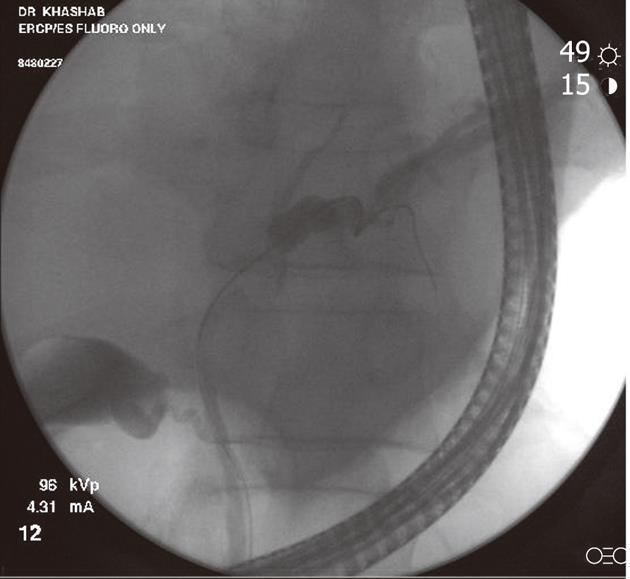
Figure 1 Fluoroscopic image during endoscopic retrograde cholangiopancreatography showing a hilar stricture with left intrahepatic biliary dilation.
Endoscopic brushing was performed and routine cytology confirmed hilar cholangiocarcinoma.
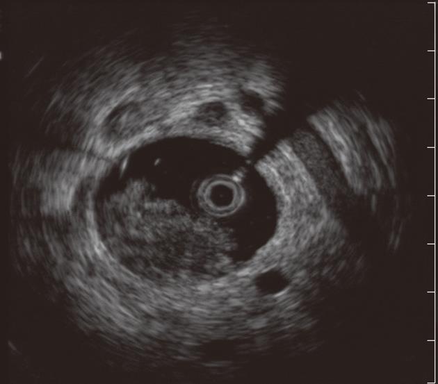
Figure 2 Intraductal ultrasound showing bile duct mass and surrounding lymph nodes.
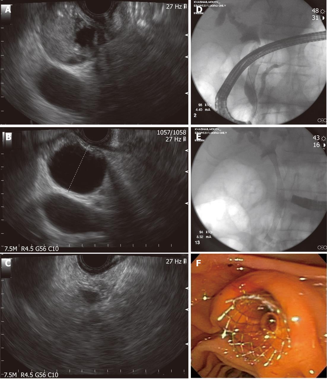
Figure 3 Endoscopic diagnosis and therapy for a bile duct mass missed on transabdominal imaging.
A: Endoscopic ultrasound showing a bile duct mass that was missed by computed tomography and magnetic resonance imaging; B: Biliary dilation was present proximal to the stenosis; C: Endoscopic ultrasound-guided fine needle aspiration was performed and was diagnostic of cholangiocarcinoma; D: Endoscopic retrograde cholangioscopy was performed during the same session and cholangiography revealed distal biliary stricture; E, F: A fully-covered metal biliary stent was placed.
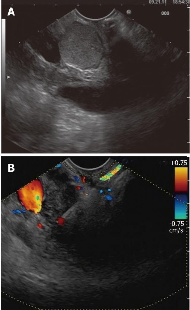
Figure 4 Endoscopic ultrasound evaluation of bile duct mass not seen on transabdominal imaging.
A: Endoscopic ultrasound demonstrating the presence of bile duct mass; B: Endoscopic ultrasound-guided fine needle aspiration was diagnostic of cholangiocarcinoma
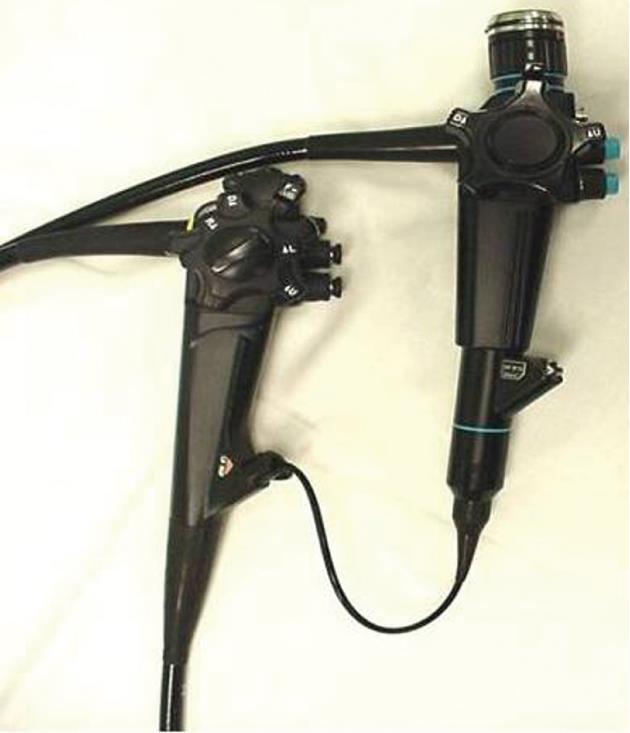
Figure 5 The “mother-baby” scope cholangioscopy system.
The main disadvantage of this system is the requirement for two endoscopists to perform the procedure.
Figure 6 Spyglass single operator cholangioscopy system.
A: SpyScope 10Fr access and delivery catheter; B: SpyGlass fiber optic probe; C: SpyBite biopsy forceps.
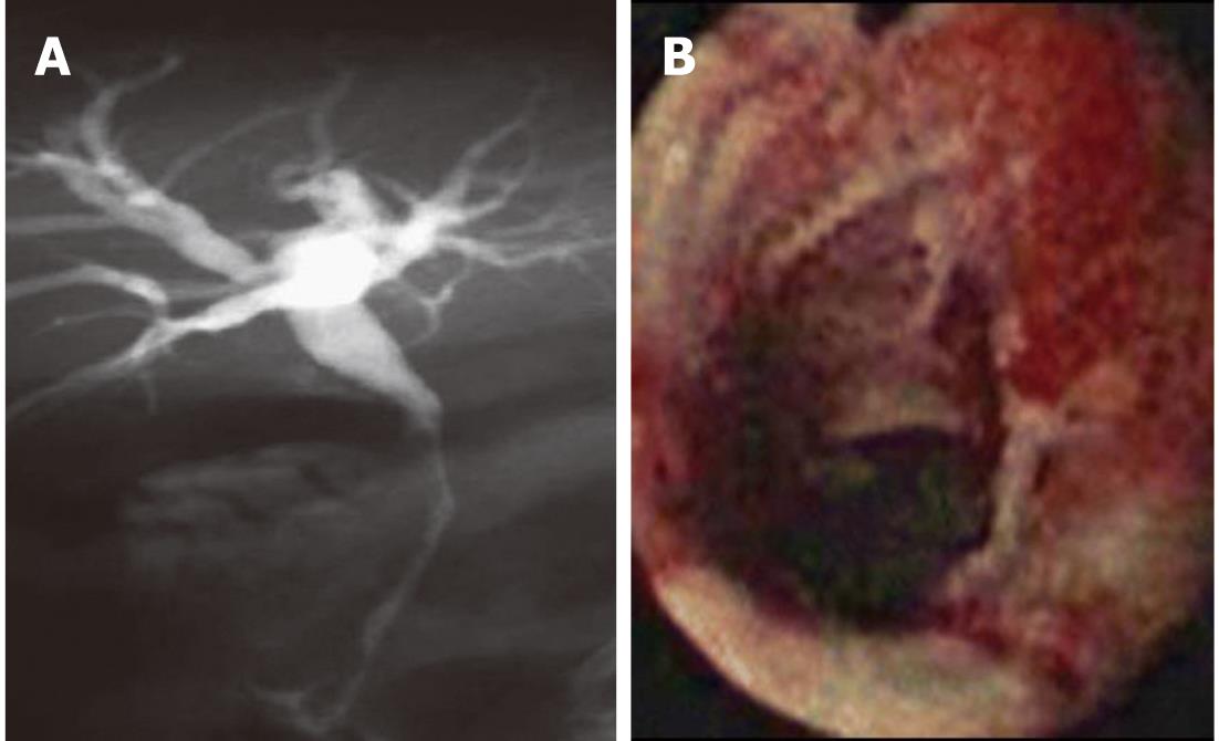
Figure 7 Single operator cholangioscopy used to obtain a diagnosis in a stricture with nondiagnostic cytology.
A: Magnetic resonance cholangiopancreatography showing a long distal biliary stricture with proximal biliary dilation. Endoscopic retrograde cholangiopancreatography with brushing was non-diagnostic; B: SpyGlass cholangioscopy revealed a malignant-appearing ulcerated biliary stricture. Spybite biopsies confirmed cholangiocarcinoma.
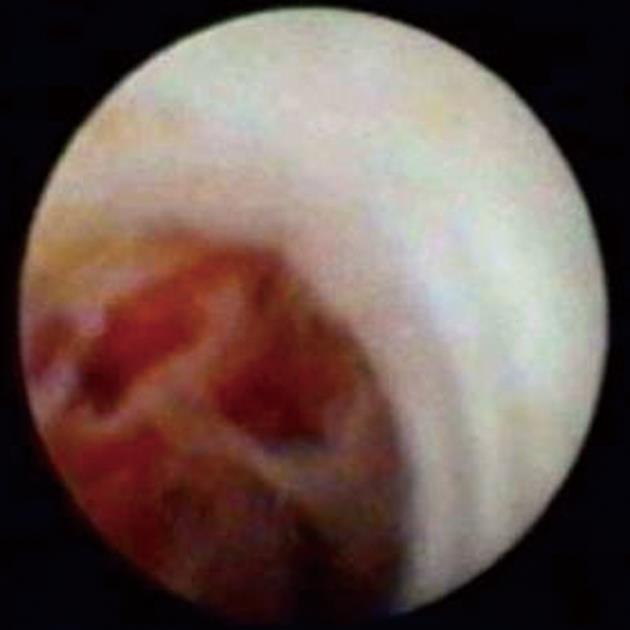
Figure 8 SpyGlass cholangioscopy revealing a bile duct mass.
This is indicative of cholangiocarcinoma.
- Citation: Victor DW, Sherman S, Karakan T, Khashab MA. Current endoscopic approach to indeterminate biliary strictures. World J Gastroenterol 2012; 18(43): 6197-6205
- URL: https://www.wjgnet.com/1007-9327/full/v18/i43/6197.htm
- DOI: https://dx.doi.org/10.3748/wjg.v18.i43.6197


