Copyright
©2012 Baishideng Publishing Group Co.
World J Gastroenterol. Nov 14, 2012; 18(42): 6160-6163
Published online Nov 14, 2012. doi: 10.3748/wjg.v18.i42.6160
Published online Nov 14, 2012. doi: 10.3748/wjg.v18.i42.6160
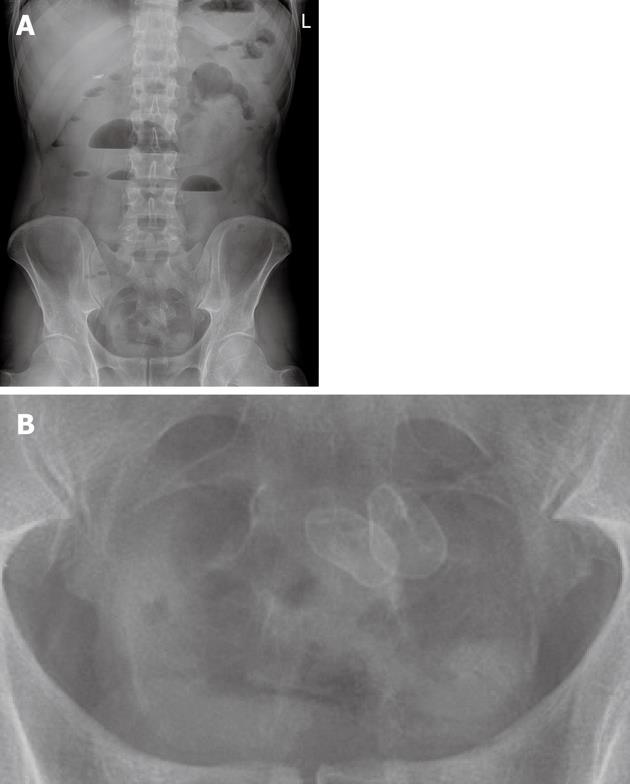
Figure 1 X-ray of the abdomen with two small radiopaque enteroliths in the lower abdomen.
A: Overall picture; B: Enlarged detail. L: Left.
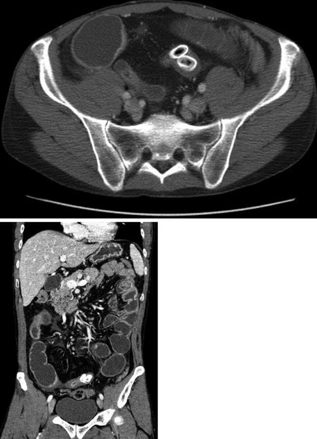
Figure 2 Computed tomography images with two radiopaque enteroliths.
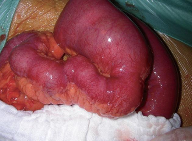
Figure 3 Intraoperative illustration of the small bowel ileus with evident stenosis and prestenotic dilation.
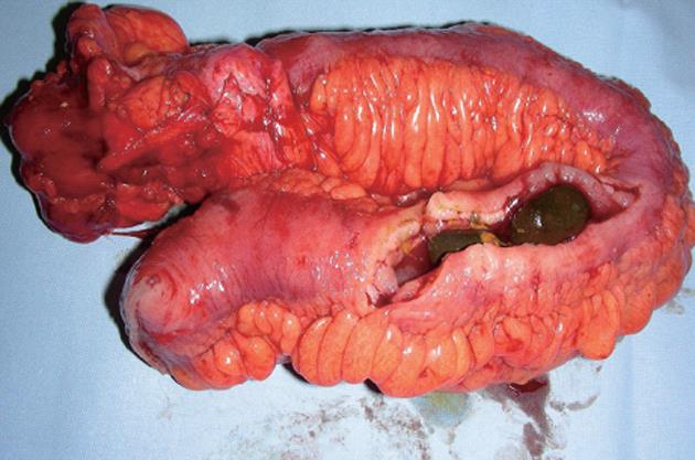
Figure 4 Resected specimen of the cecum and small bowel with two fixed enteroliths causing small bowel obstruction.
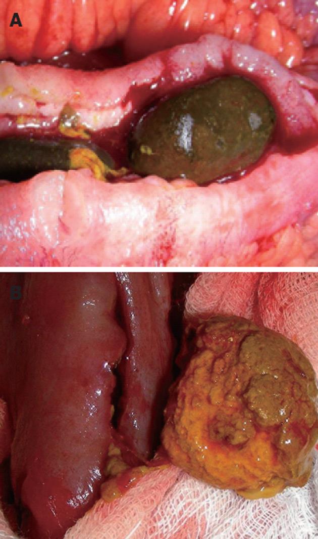
Figure 5 Typical images of enteroliths.
A: Long-standing enterolith with blank-looking polished surface; B: Emerging enterolith with a rough and fragile surface.
- Citation: Perathoner A, Kogler P, Denecke C, Pratschke J, Kafka-Ritsch R, Zitt M. Enterolithiasis-associated ileus in Crohn's disease. World J Gastroenterol 2012; 18(42): 6160-6163
- URL: https://www.wjgnet.com/1007-9327/full/v18/i42/6160.htm
- DOI: https://dx.doi.org/10.3748/wjg.v18.i42.6160


