Copyright
©2012 Baishideng Publishing Group Co.
World J Gastroenterol. Nov 7, 2012; 18(41): 5994-5998
Published online Nov 7, 2012. doi: 10.3748/wjg.v18.i41.5994
Published online Nov 7, 2012. doi: 10.3748/wjg.v18.i41.5994
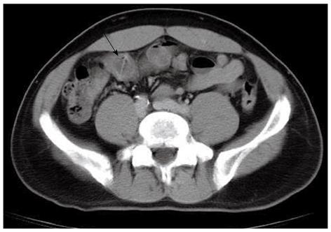
Figure 1 An unenhanced abdominal computerized tomography image reveals a 26 mm in length radiopaque linear shadow in the distal ileum lodged into the thickened intestinal wall at both ends (black arrow).
Minimal peritoneal contamination without pneumoperitoneum, or abscess formation is noted, which is consistent with signs of fish bone induced micro-perforation.
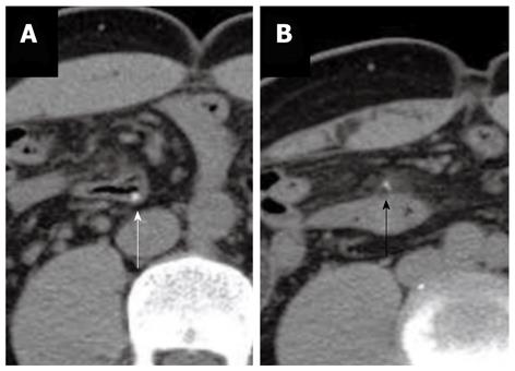
Figure 2 Two follow-up unenhanced abdominal computer tomography images, which reveal the radiopaque shadow still lodged in the intestinal segment.
The fish bone rotates and becomes parallel to the distal ileum lumen. A: Most of the fish bone is still inside the intestinal lumen (white arrow). One end of the fish bone penetrates out the intestinal wall into the mesenteric fat; B: Minimal local inflammatory infiltration contains the protruding part. No free air or abscess is noted (black arrow). The distance between these two images is 18 mm.
- Citation: Kuo CC, Jen TK, Wen CH, Liu CP, Hsiao HS, Liu YC, Chen KH. Medical treatment for a fish bone-induced ileal micro-perforation: A case report. World J Gastroenterol 2012; 18(41): 5994-5998
- URL: https://www.wjgnet.com/1007-9327/full/v18/i41/5994.htm
- DOI: https://dx.doi.org/10.3748/wjg.v18.i41.5994


