Copyright
©2012 Baishideng Publishing Group Co.
World J Gastroenterol. Nov 7, 2012; 18(41): 5990-5993
Published online Nov 7, 2012. doi: 10.3748/wjg.v18.i41.5990
Published online Nov 7, 2012. doi: 10.3748/wjg.v18.i41.5990
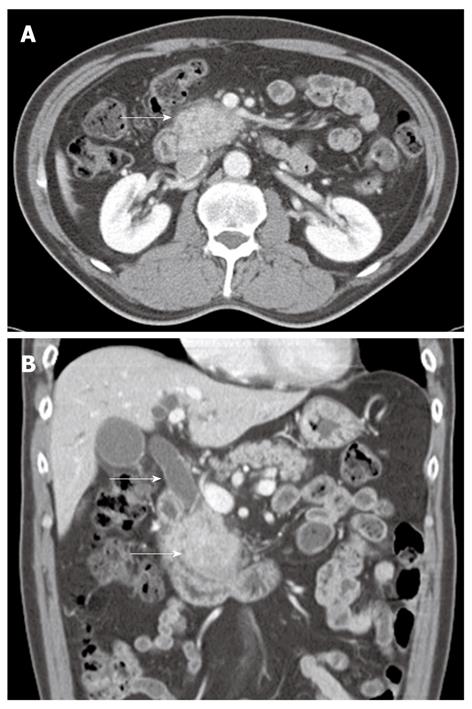
Figure 1 Initial abdominal computed tomography scan revealed an ill-defined enhancing mass-like lesion in the uncinate process of the pancreas with a dilatation of common bile duct.
A: The uncinate process of the pancreas (white arrow) measuring 5.7 cm × 3.2 cm with regional infiltrations; B: In addition to this, there is small amounts of fluid collections around the pancreas head (white arrows).
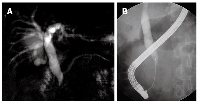
Figure 2 Magentic resonance cholangiopancreatography and endoscopic retrograde cholangiography.
A: Magentic resonance cholangiopancreatography showed moderate dilatation of the intrahepatic and common bile duct. The distal common bile duct had an abrupt narrowing. The pancreatic duct was unremarkable; B: Endoscopic retrograde cholangiography revealed a beak shaped stricture of the distal common bile duct with biliary dilatation above it.
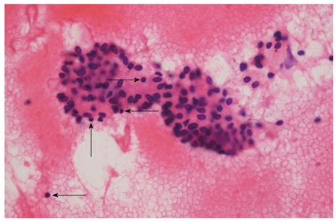
Figure 3 Photomicrograph of cytologic specimen obtained by endoscopic ultrasound-fine needle aspiration, showing lymphocytes (arrows) and irregular sheets of bland ductal epithelial cells on the bloody background (hematoxylin and eosin, × 400).
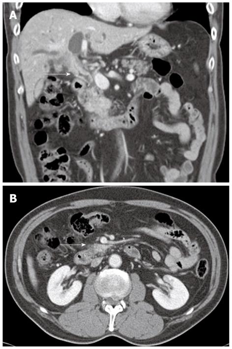
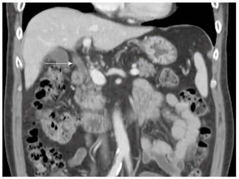
Figure 4 Follow up abdominal computed tomography scan.
A: Coronal image revealed a stenosis of the common hepatic and the proximal common bile duct (white arrow) with significant thickening and inner wall enhancement of the bile duct; B: There was no pancreatic relapse (white arrow).
Figure 5 Abdominal computed tomography scan after retreatment with prednisolone showed resolution of the thickening of the bile duct (white arrow).
- Citation: Kim JH, Chang JH, Nam SM, Lee MJ, Maeng IH, Park JY, Im YS, Kim TH, Kim CW, Han SW. Newly developed autoimmune cholangitis without relapse of autoimmune pancreatitis after discontinuing prednisolone. World J Gastroenterol 2012; 18(41): 5990-5993
- URL: https://www.wjgnet.com/1007-9327/full/v18/i41/5990.htm
- DOI: https://dx.doi.org/10.3748/wjg.v18.i41.5990


