Copyright
©2012 Baishideng Publishing Group Co.
World J Gastroenterol. Nov 7, 2012; 18(41): 5979-5981
Published online Nov 7, 2012. doi: 10.3748/wjg.v18.i41.5979
Published online Nov 7, 2012. doi: 10.3748/wjg.v18.i41.5979
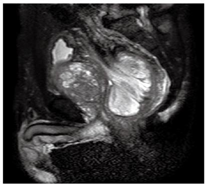
Figure 1 T2-weighted magnetic resonance image demonstrating a high-intensity mass in rectum with prostatomegaly.
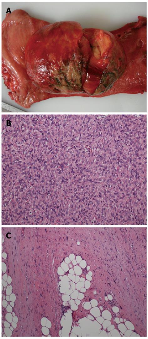
Figure 2 Dedifferentiated liposarcoma in rectum.
A: Gross picture showing a huge dedifferentiated liposarcoma in rectum; B: Dedifferentiated component identified in polypoid lesion at rectum [hematoxylin and eosin (HE), × 400]; C: Well-differentiated liposarcoma at mesorectum (HE, × 400).
- Citation: Tsuruta A, Notohara K, Park T, Itoh T. Dedifferentiated liposarcoma of the rectum: A case report. World J Gastroenterol 2012; 18(41): 5979-5981
- URL: https://www.wjgnet.com/1007-9327/full/v18/i41/5979.htm
- DOI: https://dx.doi.org/10.3748/wjg.v18.i41.5979


