Copyright
©2012 Baishideng Publishing Group Co.
World J Gastroenterol. Nov 7, 2012; 18(41): 5957-5964
Published online Nov 7, 2012. doi: 10.3748/wjg.v18.i41.5957
Published online Nov 7, 2012. doi: 10.3748/wjg.v18.i41.5957
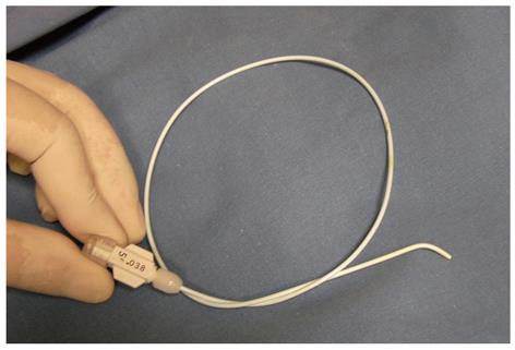
Figure 1 Kumpe catheter (5F, 40 cm).
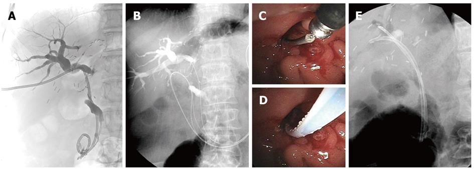
Figure 2 Guidewire technique.
A: The percutaneous transhepatic biliary drainage (PTBD) catheter was located over the anastomotic stricture into the duodenum. The angle between the right hepatic duct and the common bile duct was steep (100°); B: The 0.035 inch guidewire was inserted through the PTBD catheter, and then the PTBD catheter was removed; C: The end of the guidewire was placed outside the papilla; D: The guidewire was inserted through a bottle-top metal-tip endoscopic retrograde cholangiopancreatography (ERCP) cannula, and then the ERCP cannula was advanced into the intrahepatic bile duct; E: Two inside stents were placed over the stricture in the anterior and posterior branches of the right hepatic duct in the recipient liver.
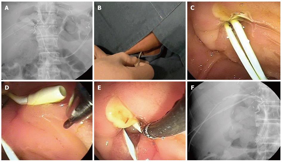
Figure 3 Kumpe catheter technique.
A: Two Kumpe (KMP) catheters were placed along the previous percutaneous transhepatic biliary drainage tracts; B, C: The KMP catheters were located out of the major ampulla in the duodenum; D, E: The KMP catheter was pulled back and rotated to approximate the slightly angulated end of the KMP catheter and the end of the endoscopic retrograde cholangiopancreatography (ERCP) cannula. Then, a preloaded guidewire in the ERCP cannula was advanced through the KMP catheter; F: Two inside stents were placed over the stricture in the anterior and posterior branches of the right hepatic duct of the recipient liver.
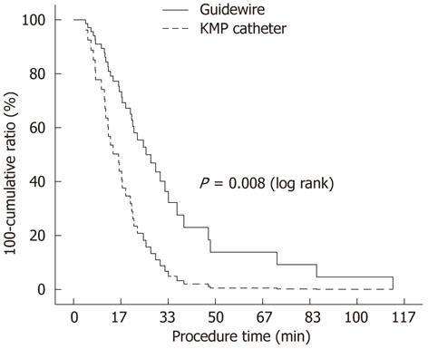
Figure 4 Cumulative probability of rendezvous procedures corresponding to procedure time.
Kumpe catheter group vs guidewire group.
- Citation: Chang JH, Lee IS, Chun HJ, Choi JY, Yoon SK, Kim DG, You YK, Choi MG, Han SW. Comparative study of rendezvous techniques in post-liver transplant biliary stricture. World J Gastroenterol 2012; 18(41): 5957-5964
- URL: https://www.wjgnet.com/1007-9327/full/v18/i41/5957.htm
- DOI: https://dx.doi.org/10.3748/wjg.v18.i41.5957


