Copyright
©2012 Baishideng Publishing Group Co.
World J Gastroenterol. Sep 28, 2012; 18(36): 5142-5144
Published online Sep 28, 2012. doi: 10.3748/wjg.v18.i36.5142
Published online Sep 28, 2012. doi: 10.3748/wjg.v18.i36.5142
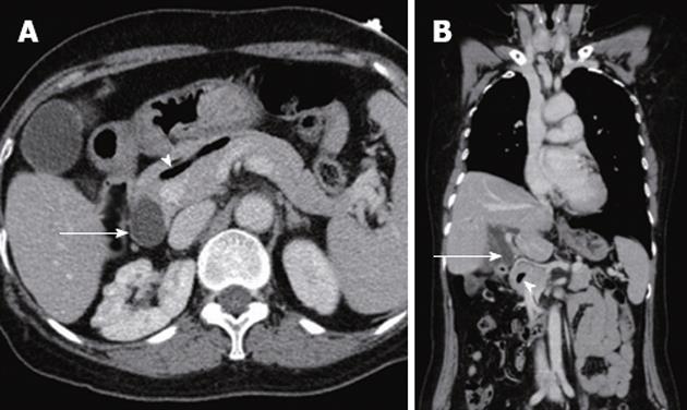
Figure 1 Computed tomography scans revealed air in the main pancreatic duct (arrowhead) and dilatation of the common bile duct (arrow).
A: Axial scan; B: Coronal scan.
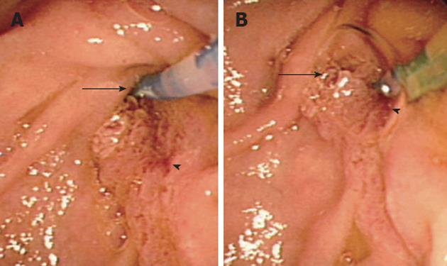
Figure 2 Duodenoscopy revealed separate biliary and pancreatic orifices.
A: A catheter inserted into the biliary orifice (arrow); B: A catheter inserted into the pancreatic orifice (arrowhead). The pancreatic orifice was patulous with some air bubbles appearing in it.
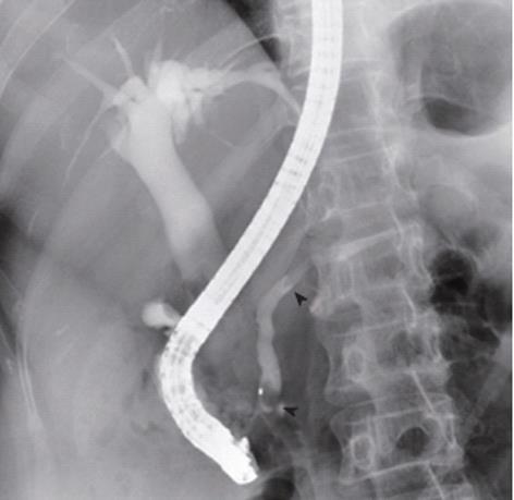
Figure 3 Endoscopic retrograde cholangiopancreatography showed dilatations of the common bile duct and pancreatic duct with some air bubbles (arrowheads), but no other abnormal lesions.
- Citation: Kim YJ, Kim HK, Cho YS, Kim SS, Chae HS, Kim SK, Kim ES, Lee SY. Air in the main pancreatic duct: A case of innocent air. World J Gastroenterol 2012; 18(36): 5142-5144
- URL: https://www.wjgnet.com/1007-9327/full/v18/i36/5142.htm
- DOI: https://dx.doi.org/10.3748/wjg.v18.i36.5142


