Copyright
©2012 Baishideng Publishing Group Co.
World J Gastroenterol. Sep 28, 2012; 18(36): 5138-5141
Published online Sep 28, 2012. doi: 10.3748/wjg.v18.i36.5138
Published online Sep 28, 2012. doi: 10.3748/wjg.v18.i36.5138
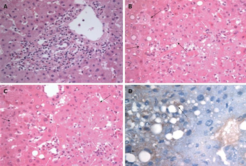
Figure 1 Histopathological analysis of a liver specimen from a patient with scrub typhus.
A: Portal inflammation showing lymphocytes and some eosinophils devoid of distinct interface hepatitis; B, C: The degeneration of individual hepatocytes (black arrows), mild ballooning changes with lobular disarray and small clusters of mononuclear cell infiltration (white arrows) identified in the lobules. Neither cholestasis nor hepatocyte apoptosis are evident (hematoxylin and eosin stain, ×400 magnification); D: Immunohistochemical staining for Orientia tsutsugamushi demonstrating scattered positive immunoreactions (arrows) in the cytoplasm of the hepatocytes (labeled streptavidin-biotin method, counterstained by hematoxylin, ×400 magnification).
- Citation: Chung JH, Lim SC, Yun NR, Shin SH, Kim CM, Kim DM. Scrub typhus hepatitis confirmed by immunohistochemical staining. World J Gastroenterol 2012; 18(36): 5138-5141
- URL: https://www.wjgnet.com/1007-9327/full/v18/i36/5138.htm
- DOI: https://dx.doi.org/10.3748/wjg.v18.i36.5138


