Copyright
©2012 Baishideng Publishing Group Co.
World J Gastroenterol. Sep 21, 2012; 18(35): 4925-4933
Published online Sep 21, 2012. doi: 10.3748/wjg.v18.i35.4925
Published online Sep 21, 2012. doi: 10.3748/wjg.v18.i35.4925
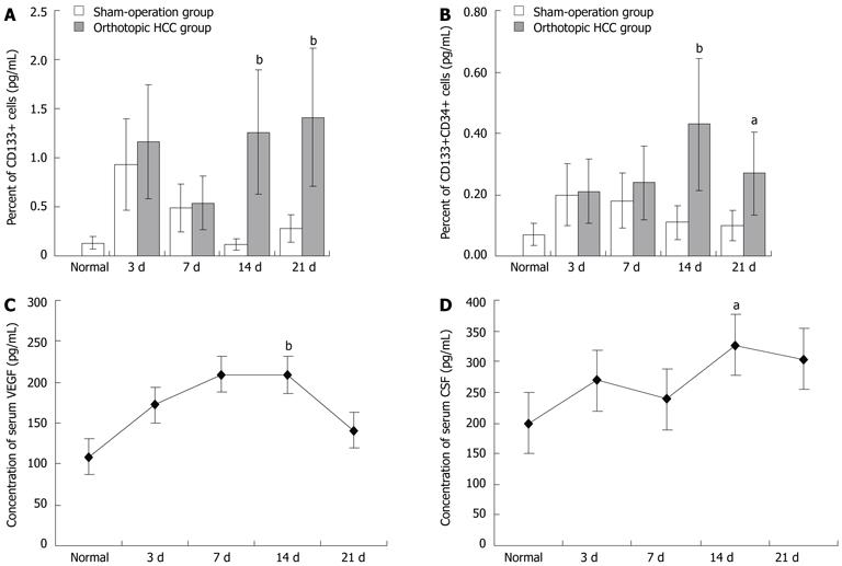
Figure 1 Mobilization of endothelial progenitor cells into circulation.
A, B: Flow cytometry analysis of circulating endothelial progenitor cells in hepatocellular carcinoma (HCC) mice (day 14, n = 7; day 21, n = 9) vs sham-operation group (day 14, n = 9; day 21, n = 11). Normal = normal mice; C, D: Dynamic changes in serum levels of vascular endothelial growth factor and colony-stimulating factor in HCC mice (n = 8). aP < 0.05, bP < 0.01 vs normal control (n = 8). CSF: Colony-stimulating factor; VEGF: Vascular endothelial growth factor.
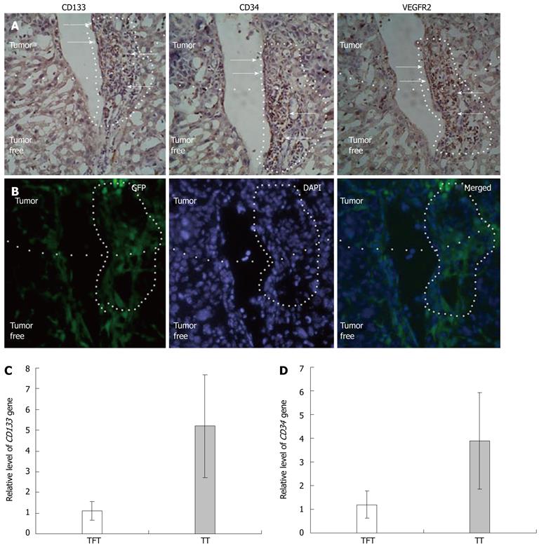
Figure 2 Recruitment of endothelial progenitor cells to hepatocellular carcinoma tissue.
A: Distribution endothelial progenitor cells (EPCs) in tumor and tumor-free tissue in four consecutive 2 μm sections (arrows: Cells expressing the indicated antigens and recruited to tumors and tumor vessels); B: Bone-marrow cells labeled by green fluorescent protein recruited to tumors and tumor vessels, occupying the same position as antigen positive cells [blue regions: Nuclei stained by DAPI (frozen section, 2 μm, magnification 200)]; C: Relative levels of CD133 mRNA in tumor tissue (n = 7) and tumor-free tissue (n = 6); D: Relative level of CD34 mRNA in tumor tissue (n = 7) and tumor-free tissue (n = 7). TFT: Tumor-free tissues; TT: Tumor tissues; DAPI: 4',6'-diamidino-2-phenylindole hydrochloride; GFP: Green fluorescent protein.
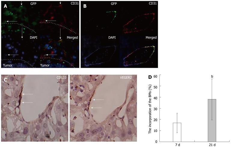
Figure 3 Bone-marrow-derived endothelial progenitor cells were found to contribute to vessel formation in hepatocellular carcinoma tissues.
A: Bone marrow derived endothelial progenitor cells (EPCs) incorporated into peritumoral vessels; B: Distribution of CD133 and VEGFR2 antigens in hepatocellular carcinoma (HCC) liver vessels (Arrows: Double-positive cells, CD133+ VEGFR2+ cells incorporating into microvessels in the liver. Magnification, 400); C: Bone-marrow (BM)-derived EPCs incorporated into hepatic veins [Red: Mature vascular endothelial cells (VECs) are marked by CD31; Green: BM derived cells expressing green fluorescent protein (GFP); Blue: Nuclei stained by DAPI; Arrows: VECs co-expressing GFP and CD31, frozen, HCC, 2 μm, magnification 400]; D: The incorporation rate of the EPCs in HCC blood vessels at 7 d vs that observed at 21 d. bP < 0.01 vs that observed at 21 d, n = 15. DAPI: 4',6'-diamidino-2-phenylindole hydrochloride.
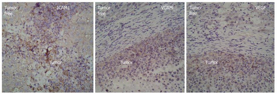
Figure 4 Expression of intercellular adhesion molecule 1, vascular adhesion molecule 1, and vascular endothelial growth factor in tumor tissues and tumor-free tissues as detected by immunohistochemistry (paraffin, liver, 2 μm, magnification, 200).
- Citation: Sun XT, Yuan XW, Zhu HT, Deng ZM, Yu DC, Zhou X, Ding YT. Endothelial precursor cells promote angiogenesis in hepatocellular carcinoma. World J Gastroenterol 2012; 18(35): 4925-4933
- URL: https://www.wjgnet.com/1007-9327/full/v18/i35/4925.htm
- DOI: https://dx.doi.org/10.3748/wjg.v18.i35.4925


