Copyright
©2012 Baishideng Publishing Group Co.
World J Gastroenterol. Sep 21, 2012; 18(35): 4898-4904
Published online Sep 21, 2012. doi: 10.3748/wjg.v18.i35.4898
Published online Sep 21, 2012. doi: 10.3748/wjg.v18.i35.4898
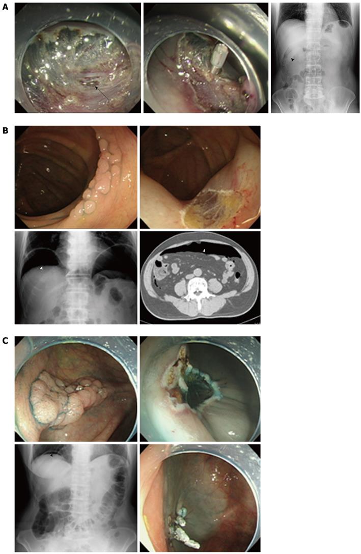
Figure 1 Representative cases of colonoscopic perforations.
A: Case 1: Perforation discovered during the therapeutic procedure and treated with clipping; B: Case 2: Perforation discovered more than 24 h later after the therapeutic procedure and treated without clipping; C: Case 3: Perforation discovered just after the therapeutic procedure and treated with suture clipping.
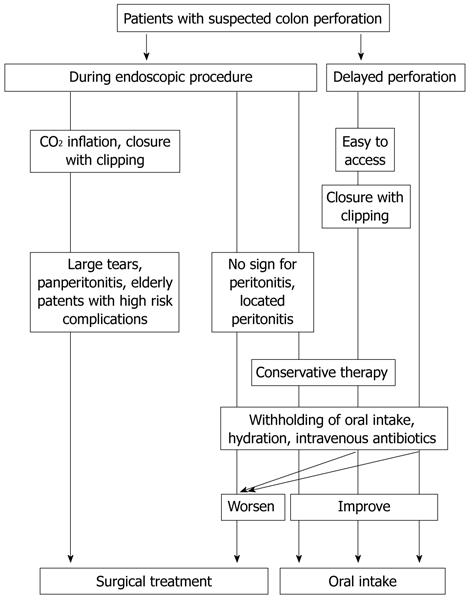
Figure 2 Trouble shooting for perforation at the local clinic.
- Citation: Sagawa T, Kakizaki S, Iizuka H, Onozato Y, Sohara N, Okamura S, Mori M. Analysis of colonoscopic perforations at a local clinic and a tertiary hospital. World J Gastroenterol 2012; 18(35): 4898-4904
- URL: https://www.wjgnet.com/1007-9327/full/v18/i35/4898.htm
- DOI: https://dx.doi.org/10.3748/wjg.v18.i35.4898


