Copyright
©2012 Baishideng Publishing Group Co.
World J Gastroenterol. Sep 14, 2012; 18(34): 4771-4780
Published online Sep 14, 2012. doi: 10.3748/wjg.v18.i34.4771
Published online Sep 14, 2012. doi: 10.3748/wjg.v18.i34.4771
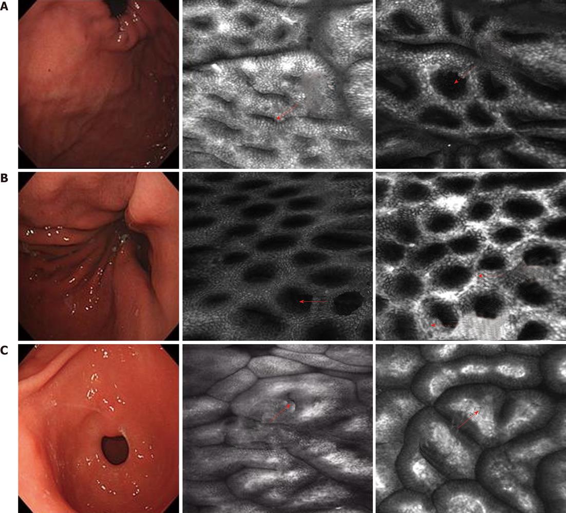
Figure 1 Routine endoscopy and confocal laser endomicroscopy images.
A: Gastric fundus: Superficial lamella gastric pits are round (solid arrow). Few pits compose one unit. The edge crack is the line type. Net-like subepithelial capillary network patterns surround the gastric pits (dash arrow); B: Gastric body: The gastric pits are round (solid arrow). Honeycomb-like subepithelial capillary network patterns surround the gastric pits (dash arrows); C: Gastric antrum: The gastric pits are the line type (solid arrow). Coil-shaped subepithelial capillary network patterns surround the gastric pits (dash arrow).
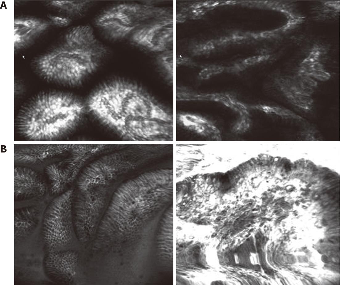
Figure 2 Confocal laser endomicroscopy images of low and high grade gastric intraepithelial neoplasia.
A: Low grade gastric intraepithelial neoplasias show that in the different sizes of gastric pits, any capillary network are thickening and circuitous; B: High grade gastric intraepithelial neoplasias show the abnormal arrangement of gastric pits. The thickening capillary network and the increasing branch present a mass shape.
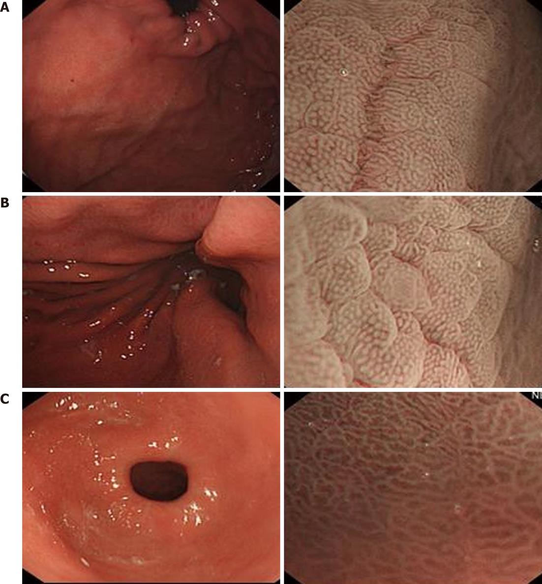
Figure 3 Routine endoscopy and intraepithelial neoplasia by narrow-band imaging images.
A: Gastric fundus; B: Gastric body; C: Gastric antrum.
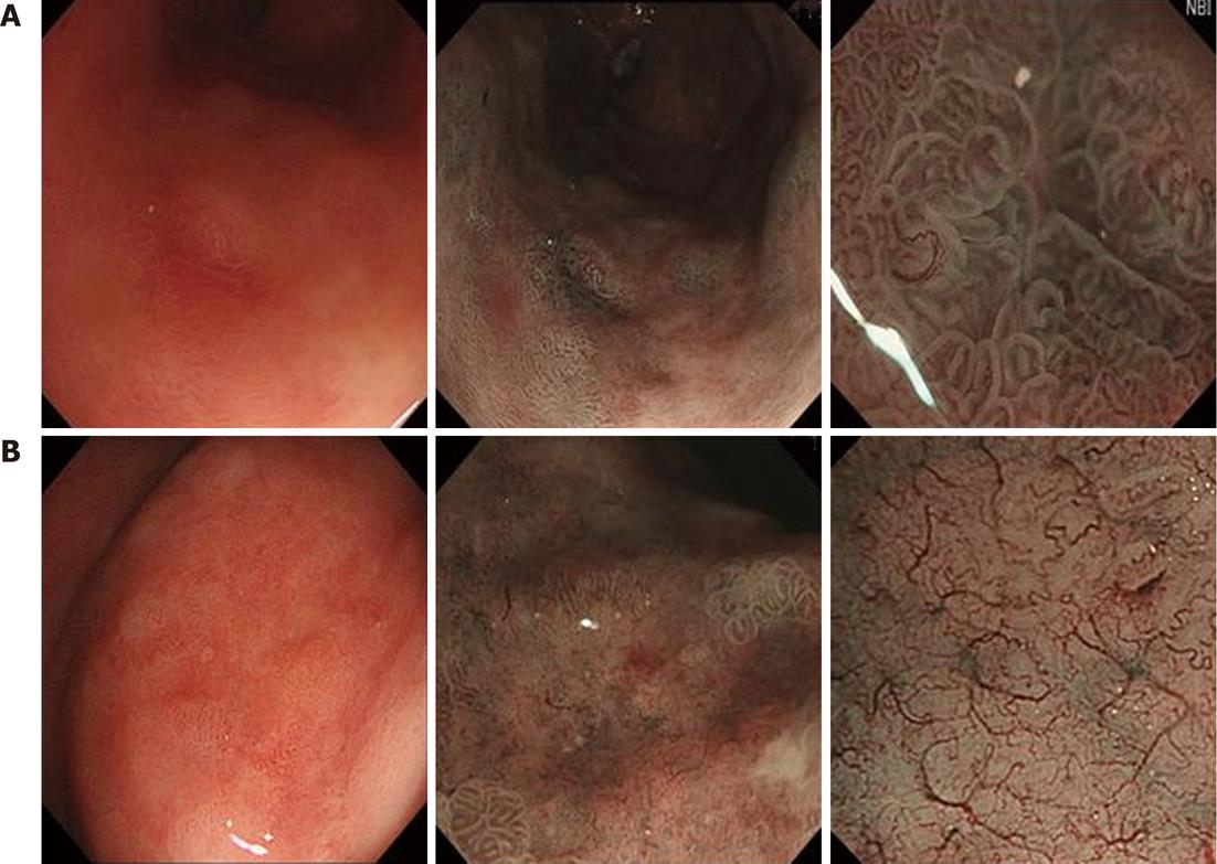
Figure 4 Routine endoscopy and narrow-band imaging images of low and high grade gastric intraepithelial neoplasia.
A: Low grade intraepithelial neoplasias present an irregular surface and microvascular pattern with a clear demarcation line; B: High grade gastric intraepithelial neoplasias present the irregular surface pattern and microvascular pattern with a demarcation line. Few areas show as flat (absent microsurface pattern) with an obscured demarcation line.
- Citation: Wang SF, Yang YS, Wei LX, Lu ZS, Guo MZ, Huang J, Peng LH, Sun G, Ling-Hu EQ, Meng JY. Diagnosis of gastric intraepithelial neoplasia by narrow-band imaging and confocal laser endomicroscopy. World J Gastroenterol 2012; 18(34): 4771-4780
- URL: https://www.wjgnet.com/1007-9327/full/v18/i34/4771.htm
- DOI: https://dx.doi.org/10.3748/wjg.v18.i34.4771


