Copyright
©2012 Baishideng Publishing Group Co.
World J Gastroenterol. Aug 28, 2012; 18(32): 4335-4341
Published online Aug 28, 2012. doi: 10.3748/wjg.v18.i32.4335
Published online Aug 28, 2012. doi: 10.3748/wjg.v18.i32.4335
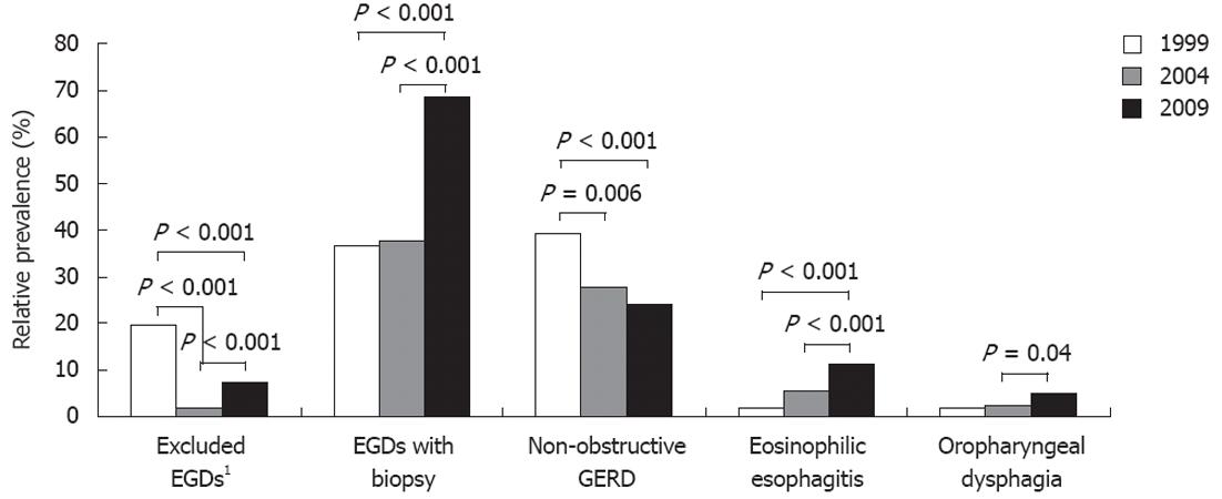
Figure 1 Trends in relative prevalence of dysphagia etiologies from 1999-2009.
GERD: Gastroesophageal reflux disease. 1Including cricopharyngeal bar, globus, functional dysphagia, diverticulum, scleroderma, and epidermolysis bullosa with proximal stricture.
Figure 2 Selected post-hoc Tukey analysis.
1Expressed as percentage of total upper endoscopies for dysphagia in the given year. GERD: Gastroesophageal reflux disease; EGD: Upper endoscopy.
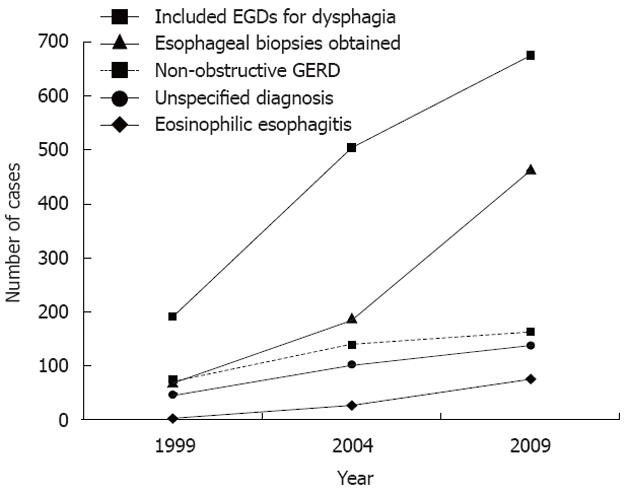
Figure 3 Trends in upper endoscopy volume, esophageal biopsies, and dysphagia diagnoses.
GERD: Gastroesophageal reflux disease; EGDs: Upper endoscopies.
- Citation: Kidambi T, Toto E, Ho N, Taft T, Hirano I. Temporal trends in the relative prevalence of dysphagia etiologies from 1999-2009. World J Gastroenterol 2012; 18(32): 4335-4341
- URL: https://www.wjgnet.com/1007-9327/full/v18/i32/4335.htm
- DOI: https://dx.doi.org/10.3748/wjg.v18.i32.4335


