Copyright
©2012 Baishideng Publishing Group Co.
World J Gastroenterol. Aug 28, 2012; 18(32): 4317-4322
Published online Aug 28, 2012. doi: 10.3748/wjg.v18.i32.4317
Published online Aug 28, 2012. doi: 10.3748/wjg.v18.i32.4317
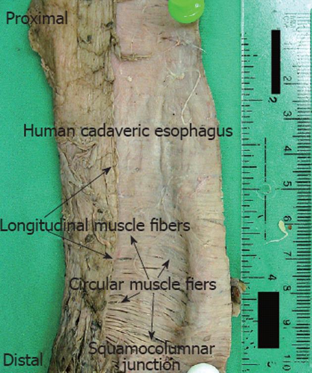
Figure 1 A photograph of a dissected cadaveric esophagus.
The esophagus is cut longitudinally along the longitudinal muscle fibers after dissecting the serosa. The mucosa and sub mucosa were later dissected. The esophagus was later folded back so that the longitudinal fibers were on top of the circular muscle fibers. Measurements of the angle of the circular smooth muscle fibers with respect to the longitudinal smooth muscle fibers were made starting from the squamocolumnar junction.
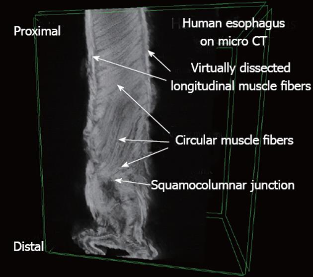
Figure 2 A human esophagus obtained using micro-computed tomography.
The longitudinal muscle fibers were virtually dissected to expose the underlying circular muscle fibers. Measurements were made starting from the squamocolumnar junction.
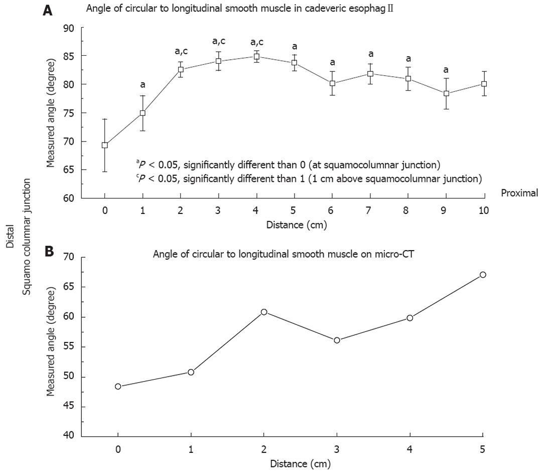
Figure 3 The average angle of the circular smooth muscle with respect to the longitudinal smooth muscle.
A: On cadaveric esophagi. The x-axis represents the distance from the squamocolumnar junction with 0 being the squamocolumnar junction. At 90 degrees the circular smooth muscle and longitudinal smooth muscle are perpendicular to each other. Therefore the smaller the angle the greater the deviation from the perpendicular and the greater the resulting shortening during peristaltic contraction; B: Measured using micro computed tomography. The x-axis represents the distance from the squamocolumnar junction with 0 being the squamocolumnar junction. CT: Computed tomography.
- Citation: Vegesna AK, Chuang KY, Besetty R, Phillips SJ, Braverman AS, Barbe MF, Ruggieri MR, Miller LS. Circular smooth muscle contributes to esophageal shortening during peristalsis. World J Gastroenterol 2012; 18(32): 4317-4322
- URL: https://www.wjgnet.com/1007-9327/full/v18/i32/4317.htm
- DOI: https://dx.doi.org/10.3748/wjg.v18.i32.4317


