Copyright
©2012 Baishideng Publishing Group Co.
World J Gastroenterol. Aug 7, 2012; 18(29): 3896-3903
Published online Aug 7, 2012. doi: 10.3748/wjg.v18.i29.3896
Published online Aug 7, 2012. doi: 10.3748/wjg.v18.i29.3896
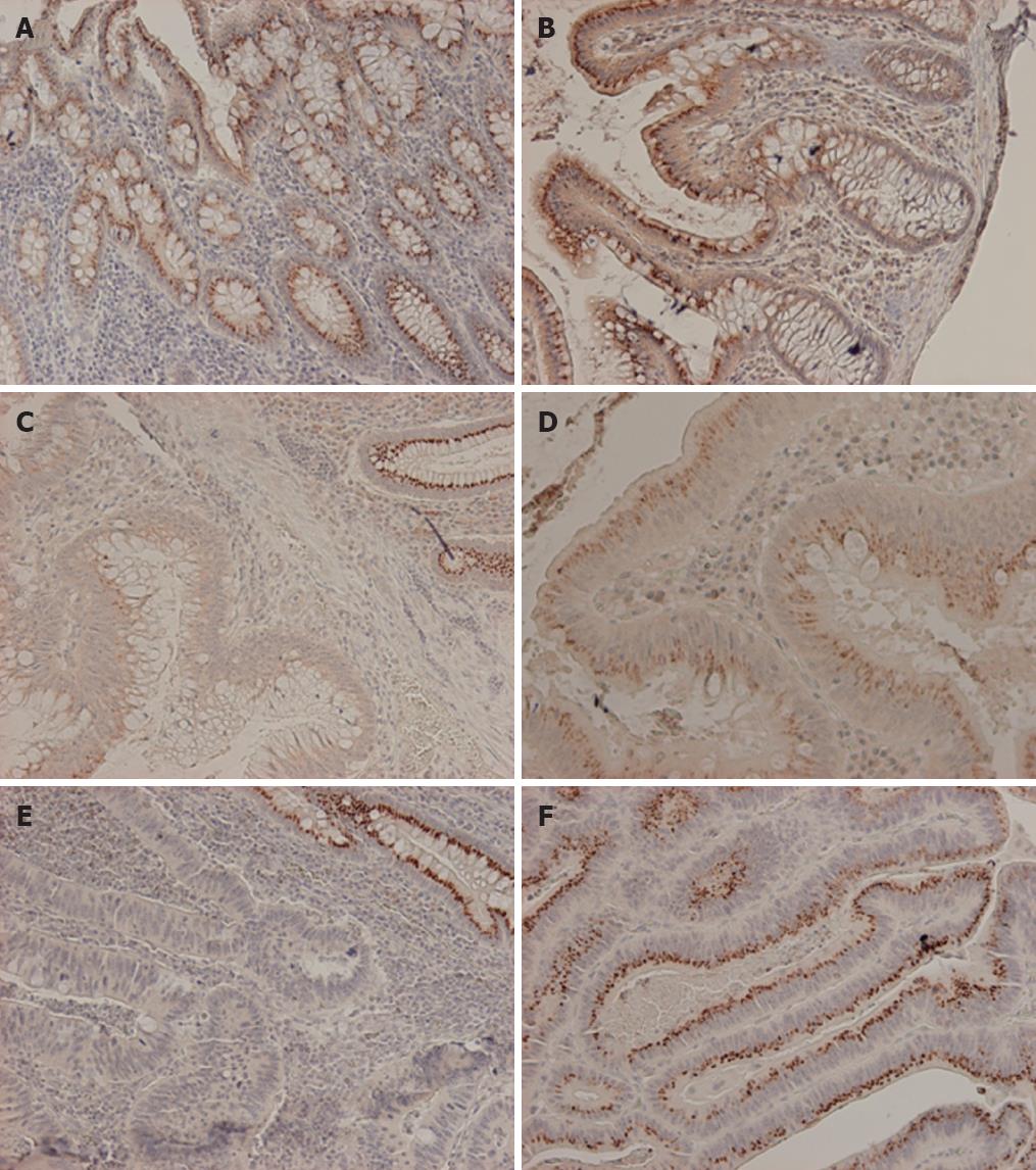
Figure 1 Epidermal growth factor receptor pathway substrate 8 immunohistochemistry.
A: Normal colon mucosa with dot-like, supranuclear expression; B: Normal colon mucosa with clear gradient of expression stronger at the luminal aspect compared to the crypt bases; C: Marked reduction to complete loss of epidermal growth factor receptor pathway substrate 8 (EPS8) expression in adenoma (lower left) compared to normal mucosa (upper right); D: EPS8 positive adenoma; E: Marked reduction to complete loss of EPS8 expression in carcinoma (lower left) compared to normal mucosa (upper right); F: EPS8 positive carcinoma.
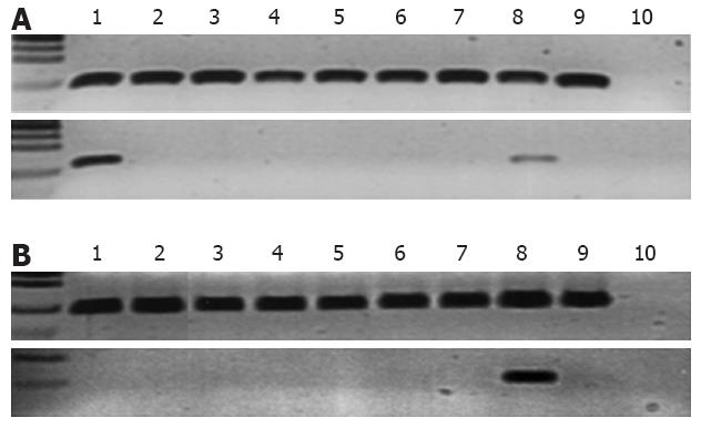
Figure 2 Methylation-specific polymerase chain reaction assays for epidermal growth factor receptor pathway substrate 8 gene.
A: Cell lines analysis; upper panel is the unmethylated (UF1 + UR1) reaction and lower panel is the methylated (MF1 + MR1) reaction. Sample order in both panels from left to right is 1, RKO; 2, LoVo; 3, LIM1215; 4, HCA7; 5, HCT116; 6, KM12; 7, HCT15; 8, TK6 (a lymphoblastoid cell line); 9, unmethylated (negative) control; 10, water; B: Primary uncultured colon cancer analysis; upper panel is the unmethylated reaction and lower panel is the methylated reaction (MF3 + MR3). Sample order in both panels from left to right is 1-7, uncultured colon cancer specimens; 8, RKO (used as methylated control); 9, unmethylated (negative) control; 10, water.
- Citation: Abdel-Rahman WM, Ruosaari S, Knuutila S, Peltomäki P. Differential roles of EPS8 in carcinogenesis: Loss of protein expression in a subset of colorectal carcinoma and adenoma. World J Gastroenterol 2012; 18(29): 3896-3903
- URL: https://www.wjgnet.com/1007-9327/full/v18/i29/3896.htm
- DOI: https://dx.doi.org/10.3748/wjg.v18.i29.3896


