Copyright
©2012 Baishideng Publishing Group Co.
World J Gastroenterol. Jul 14, 2012; 18(26): 3465-3471
Published online Jul 14, 2012. doi: 10.3748/wjg.v18.i26.3465
Published online Jul 14, 2012. doi: 10.3748/wjg.v18.i26.3465
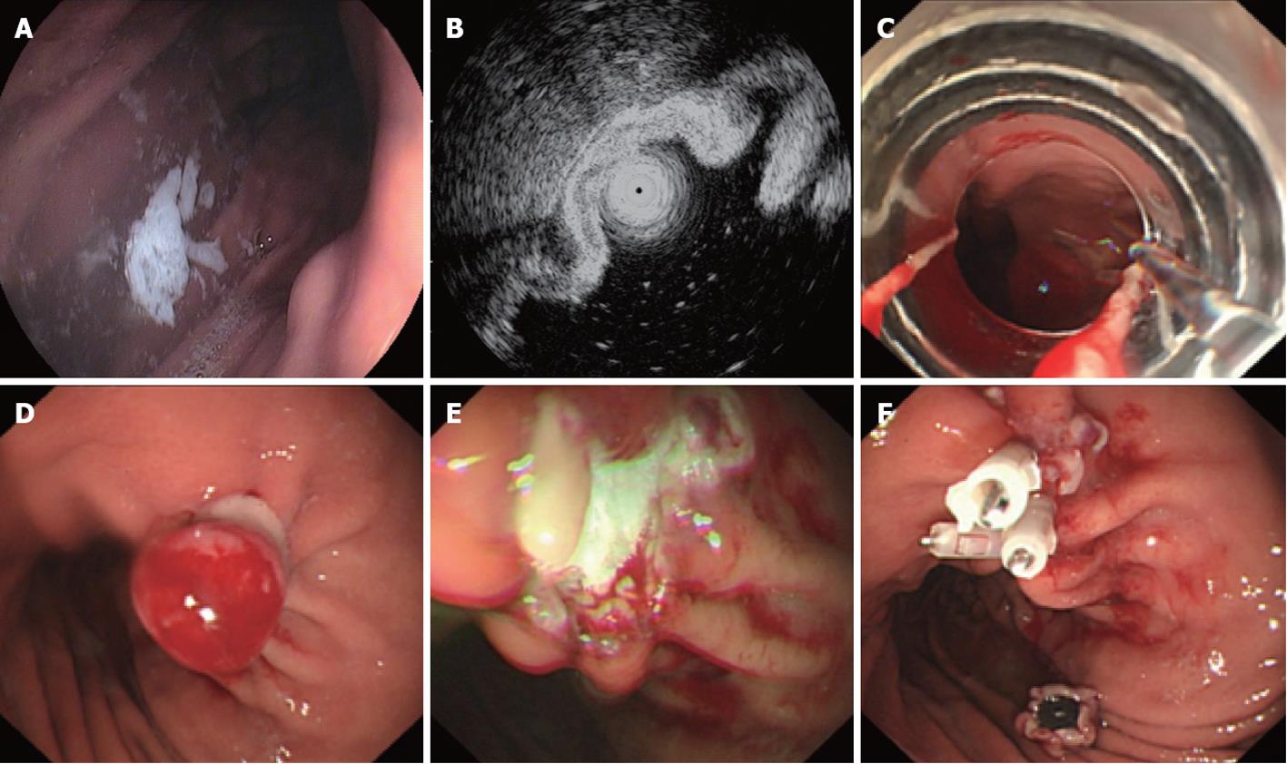
Figure 1 Endoscopic ligation and resection treatment for a gastric stromal tumor less than 1.
2 cm in size originating from the muscularis propria. A: Submucosa lesion at the posterior wall of the gastric corpus; B: Endoscopic ultrasound shows that the lesion originates from the muscularis propria; C: COOK ligator aimed at the lesion, ready to ligate; D: The ligated stromal tumor was in the shape of a polypoid with deuto-stem; E: A snare was used to cut the tumor above the rubber band; F: The wound surface was closed with metal clips.
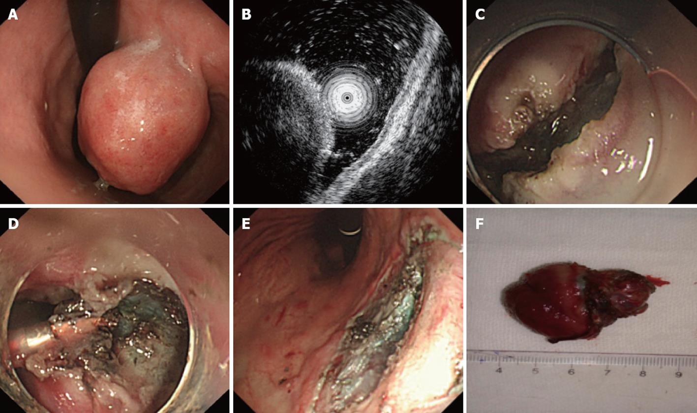
Figure 2 Endoscopic submucosal excavation treatment for a gastric stromal tumor originating from the muscularis propria that is larger than 1.
2 cm. A: Submucosa lesion at the gastric corpus; B: Endoscopic ultrasound shows that the lesion originates from the muscularis propria; C: The mucosa of the stromal tumor was cut after submucosal injection; D: Dissection with an IT knife; E: The excavated wound surface, showing that no perforation occurred; F: The resected stromal tumor (4 cm in size).
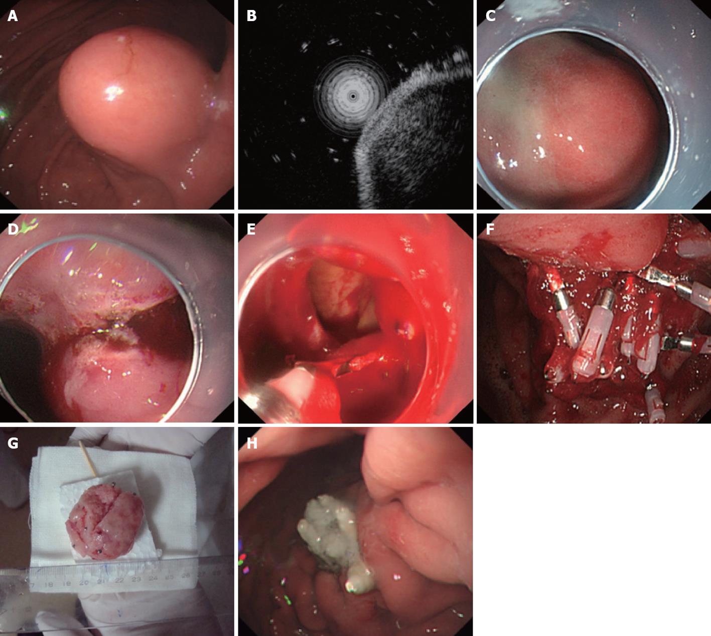
Figure 3 Endoscopic full-thickness resection treatment for a gastric stromal tumor originating from the muscularis propria larger than 1.
2 cm. A: Submucosa lesion at the gastric corpus; B: Endoscopic ultrasound shows that the lesion originates from the muscularis propria; C: Submucosal injection of the mixture of indicarminum, adrenalin and physiological saline; D: Dissection with an IT knife; E: “Artificial” perforation after gastric stromal tumor resection, closed with metal clips; F: Many clips used to close the wound defect; G: The resected tumor without mucosa (5 cm in size); H: The perforation healed 9 d after endoscopic full-thickness resection.
- Citation: Huang LY, Cui J, Liu YX, Wu CR, Yi DL. Endoscopic therapy for gastric stromal tumors originating from the muscularis propria. World J Gastroenterol 2012; 18(26): 3465-3471
- URL: https://www.wjgnet.com/1007-9327/full/v18/i26/3465.htm
- DOI: https://dx.doi.org/10.3748/wjg.v18.i26.3465


