Copyright
©2012 Baishideng Publishing Group Co.
World J Gastroenterol. Jul 14, 2012; 18(26): 3409-3414
Published online Jul 14, 2012. doi: 10.3748/wjg.v18.i26.3409
Published online Jul 14, 2012. doi: 10.3748/wjg.v18.i26.3409
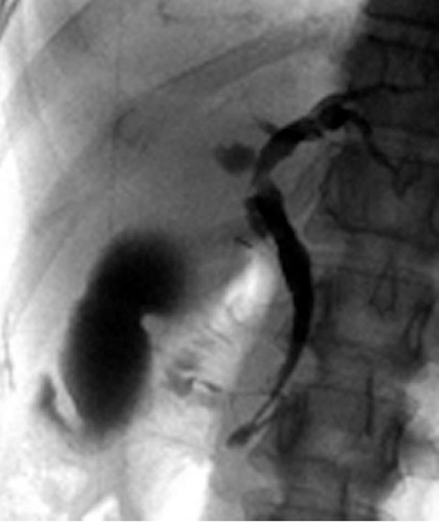
Figure 1 Endoscopic retrograde cholangiopancreatography of a patient with pancreaticobiliary maljunction without biliary dilatation showing a long common channel and deformity with fuzzy irregularity of the gallbladder.
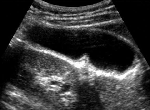
Figure 2 Abdominal ultrasound of a patient with pancreaticobiliary maljunction without biliary dilatation showing uniform smooth thickness of the gallbladder wall.
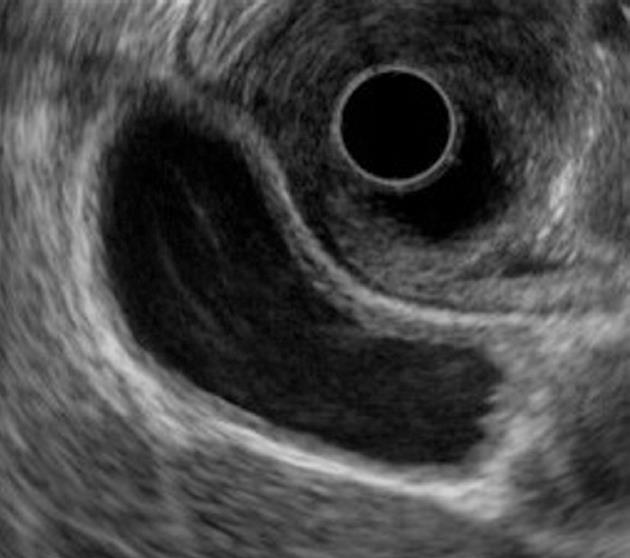
Figure 3 Endoscopic ultrasonography of a patient with pancreaticobiliary maljunction without biliary dilatation showing thickening of the inner hypoechoic layer and the outer hyperechoic layer.
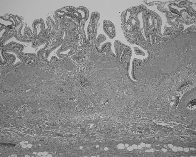
Figure 4 Histological findings of the gallbladder of a patient with pancreaticobiliary maljunction without biliary dilatation showing wall thickness composed of epithelial hyperplasia, hypertrophic muscular layer, and subserosal fibrosis.
- Citation: Takuma K, Kamisawa T, Tabata T, Hara S, Kuruma S, Inaba Y, Kurata M, Honda G, Tsuruta K, Horiguchi SI, Igarashi Y. Importance of early diagnosis of pancreaticobiliary maljunction without biliary dilatation. World J Gastroenterol 2012; 18(26): 3409-3414
- URL: https://www.wjgnet.com/1007-9327/full/v18/i26/3409.htm
- DOI: https://dx.doi.org/10.3748/wjg.v18.i26.3409


