Copyright
©2012 Baishideng Publishing Group Co.
World J Gastroenterol. Jul 7, 2012; 18(25): 3327-3330
Published online Jul 7, 2012. doi: 10.3748/wjg.v18.i25.3327
Published online Jul 7, 2012. doi: 10.3748/wjg.v18.i25.3327
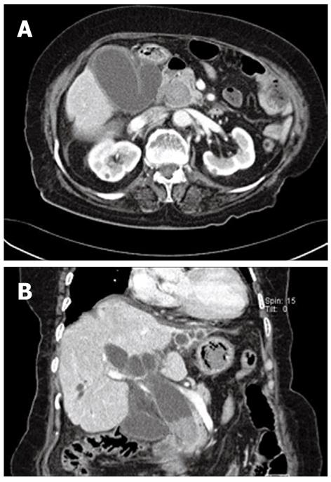
Figure 1 Computed tomogram revealed a huge stone in the distal common bile duct.
Marked dilation of the gallbladder and bile duct was also seen. A: Axial view; B: Coronal view.
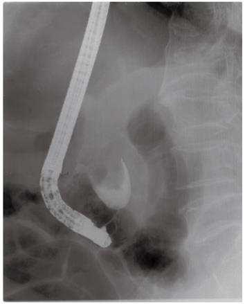
Figure 2 Endoscopic retrograde cholangiopancreatogram showed a large filling defect measuring 4.
0 cm × 3.2 cm. The proximal portion of bile duct was not opacified.
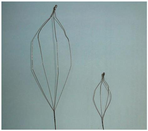
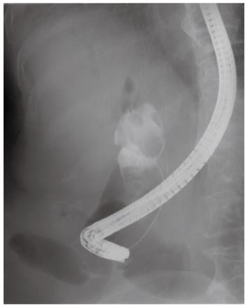
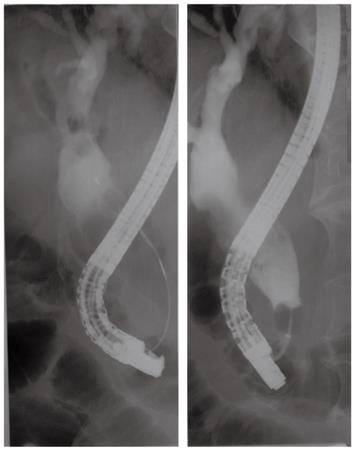
Figure 3 Basket for gastric bezoar (left) is larger than a conventional basket used for mechanical lithotripsy (right).
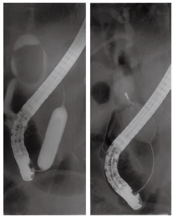
Figure 4 Endoscopic balloon dilation using a large balloon and mechanical lithotripsy.
The balloon was inflated gradually to a diameter of 15 mm across the papilla and the ballooning was sustained for 60 s, and the bezoar basket captured the stone completely. The stone was fragmented by mechanical lithotripsy using a bezoar basket.
Figure 5 Follow-up endoscopic retrograde cholangiopancreatogram disclosed remnant stone fragments.
Figure 6 Remnant stone fragments were extracted by the basket and balloon occluded cholangiogram showed complete duct clearance.
- Citation: Chung HJ, Jeong S, Lee DH, Lee JI, Lee JW, Bang BW, Kwon KS, Kim HK, Shin YW, Kim YS. Giant choledocholithiasis treated by mechanical lithotripsy using a gastric bezoar basket. World J Gastroenterol 2012; 18(25): 3327-3330
- URL: https://www.wjgnet.com/1007-9327/full/v18/i25/3327.htm
- DOI: https://dx.doi.org/10.3748/wjg.v18.i25.3327


