Copyright
©2012 Baishideng Publishing Group Co.
World J Gastroenterol. Jul 7, 2012; 18(25): 3288-3295
Published online Jul 7, 2012. doi: 10.3748/wjg.v18.i25.3288
Published online Jul 7, 2012. doi: 10.3748/wjg.v18.i25.3288
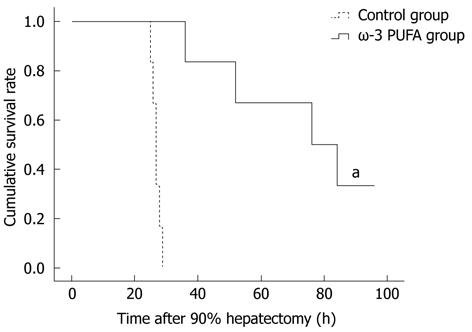
Figure 1 Survival rates in the control and the omega-3 polyunsaturated fatty acid groups.
All rats from the control group died within 30 h after hepatectomy. In contrast, in the omega-3 polyunsaturated fatty acid (ω-3 PUFA) group, 20 (100%) and 4 (20%) rats were alive at 30 h and 1 wk after PH, respectively. aP < 0.05 vs normal group.
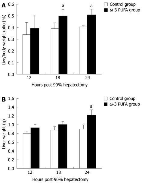
Figure 2 Change in liver weight/body weight ratios and liver weights after partial hepatectomy.
Liver weight/body weight ratios (A) and liver weights (B) in the control and the omega-3 polyunsaturated fatty acid (ω-3 PUFA) group at 12 h, 18 h and 24 h post-hepatectomy. Data are expressed as mean ± SD. n = 6 in each group. aP < 0.05 vs normal group.
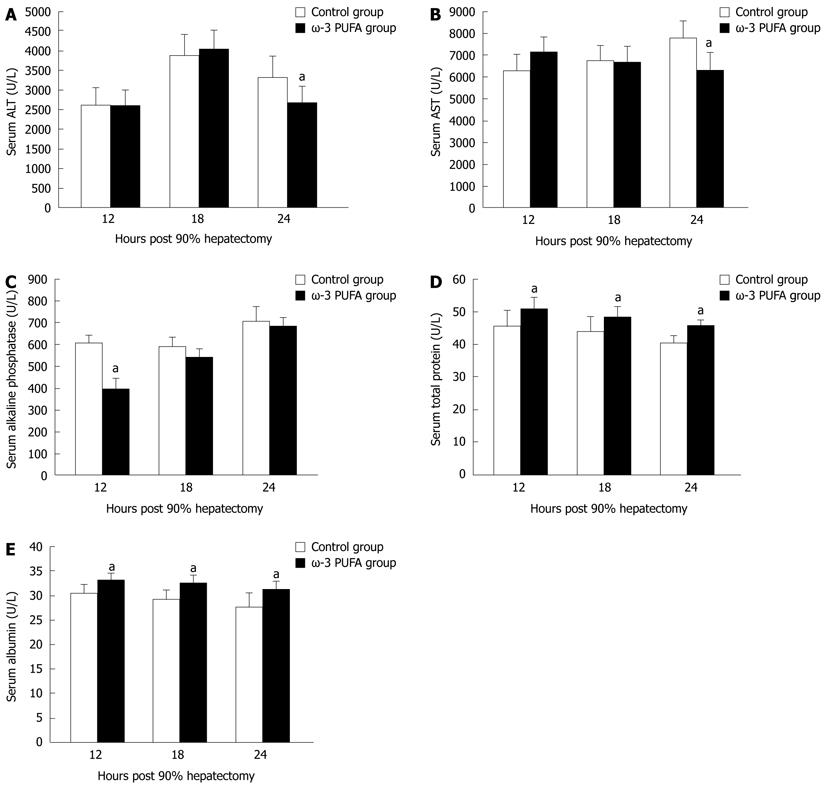
Figure 3 Serum parameters of the control and the omega-3 polyunsaturated fatty acid groups.
Serum alanine aminotransferase (ALT) (A), aspartate aminotransferase (AST) (B), alkaline phosphatase (C), total protein (D) and albumin (E) levels at 12 h, 18 h and 24 h post-hepatectomy. n = 6 in each group. aP < 0.05 vs control group. ω-3 PUFA: Omega-3 polyunsaturated fatty acid.
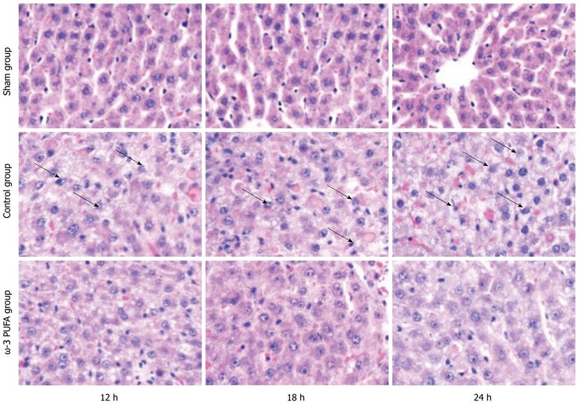
Figure 4 Histopathological examination of rats livers.
Swelling and balloon degeneration of hepatocytes were observed at all time points after partial hepatectomy (PH) in the control group (hematoxylin and eosin staining, original magnification × 400). By contrast, no significant hepatocyte swelling or balloon degeneration were observed at 12 h after PH in the omega-3 polyunsaturated fatty acid (ω-3 PUFA) group (arrow). Hepatocytes are normal and structure of liver lobules was obvious at 24 h after PH.
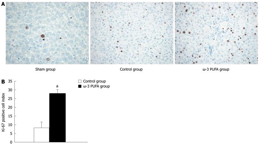
Figure 5 Change in Ki-67-positive cells after partial hepatectomy.
A: The number of Ki-67-positive cells in the liver tissue 24 h after 90% partial hepatectomy (PH) was much higher in the omega-3 polyunsaturated fatty acid (ω-3 PUFA) group; B: Compared with control group, Ki-67-positive cells were significantly increased in the ω-3 PUFA group 24 h after PH. n = 6 in each group. aP < 0.05 vs control group.
Figure 6 Change in the structure of sinusoidal endothelial cell after partial hepatectomy.
A: Normal structure of liver tissue; B: Space of Disse was enlarged and the structure of sinusoidal endothelial cell (SEC) was severely damaged at 24 h after partial hepatectomy (PH) in the control group (arrow); C: Compared with control group, the SEC structure and space of Disse were greatly restored at 24 h after PH in the omega-3 polyunsaturated fatty acid group.
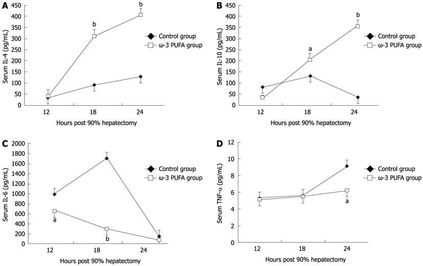
Figure 7 Change in serum cytokines.
Cytokines interleukin (IL)-4 (A) and IL-10 (B) significantly increased at 24 h in the omega-3 polyunsaturated fatty acid (ω-3 PUFA) group after partial hepatectomy, while IL-6 (C), and tumor necrosis factor (TNF)-α (D) significantly decreased at 18 h and 24 h after operation in the ω-3 PUFA group. aP < 0.05, bP < 0.01 vs control group.
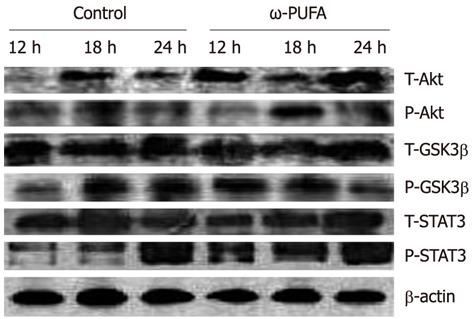
Figure 8 Phosphorylation of activation of protein kinase, signal transducer and activator of transcription 3, glycogen synthase kinase-3β.
Phosphorylation of activation of protein kinase B (Akt), signal transducer and activator of transcription 3 (STAT3), glycogen synthase kinase (GSK)-3β in the control and the omega-3 polyunsaturated fatty acid (ω-3 PUFA) groups at 12 h, 18 h and 24 h after partial hepatectomy. Liver lysates at 12 h, 18 h and 24 h time points in the quantity of 25 μg per lane were subjected to sodium dodecyl sulfate-polyacrylamide gel electrophoresis (SDS-PAGE). Proteins were transferred to polyvinylidene fluoride membrane and then incubated with specific antibodies. There was stronger phosphorylation of Akt at the 18 h time points and earlier phosphorylation of GSK3β at the 12 h time points compared to the control group. Phosphorylation of STAT3 was stronger and lasted longer at all time points in the ω-3 PUFA group.
- Citation: Qiu YD, Wang S, Yang Y, Yan XP. Omega-3 polyunsaturated fatty acids promote liver regeneration after 90% hepatectomy in rats. World J Gastroenterol 2012; 18(25): 3288-3295
- URL: https://www.wjgnet.com/1007-9327/full/v18/i25/3288.htm
- DOI: https://dx.doi.org/10.3748/wjg.v18.i25.3288


