Copyright
©2012 Baishideng Publishing Group Co.
World J Gastroenterol. Jun 28, 2012; 18(24): 3173-3176
Published online Jun 28, 2012. doi: 10.3748/wjg.v18.i24.3173
Published online Jun 28, 2012. doi: 10.3748/wjg.v18.i24.3173
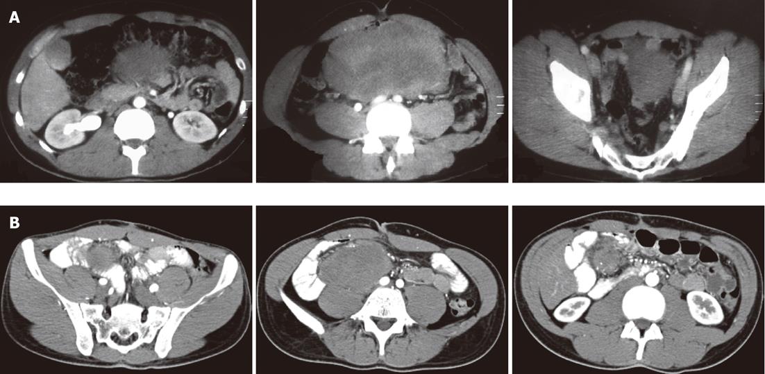
Figure 1 Abdominal computed tomography of desmoid tumour.
A: A solid large mass (22 cm × 10 cm × 15 cm) with density equal to that of soft tissue; B: A perianastomotic solid mass (13 cm × 11 cm × 10 cm) arising from the mesentery.
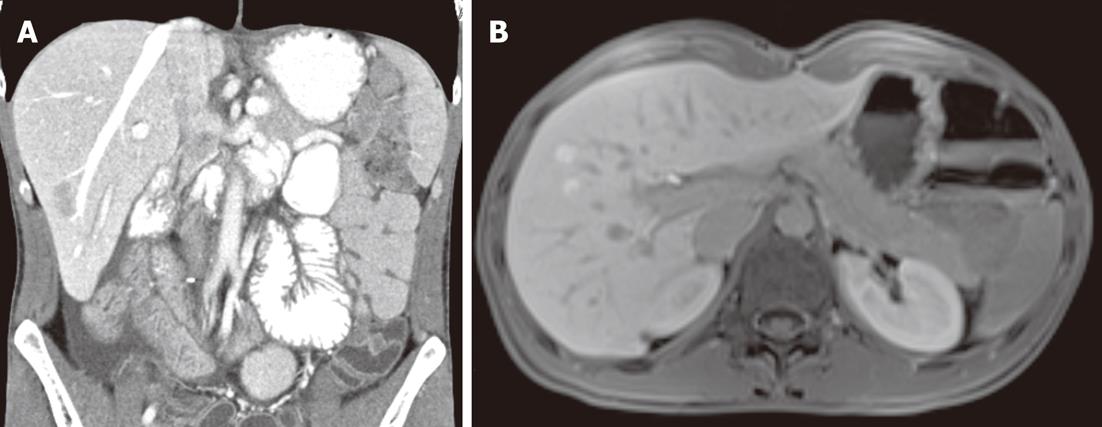
Figure 2 Fatty liver.
Abdominal computed tomography (A) and magnetic resonance imaging (B) demonstrated liver alterations with no distinguishing marks.
- Citation: Felice FD, Musio D, Caiazzo R, Dipalma B, Grapulin L, Semproni CP, Tombolini V. An unusual case of fatty liver in a patient with desmoid tumor. World J Gastroenterol 2012; 18(24): 3173-3176
- URL: https://www.wjgnet.com/1007-9327/full/v18/i24/3173.htm
- DOI: https://dx.doi.org/10.3748/wjg.v18.i24.3173


