Copyright
©2012 Baishideng Publishing Group Co.
World J Gastroenterol. Jun 28, 2012; 18(24): 3112-3118
Published online Jun 28, 2012. doi: 10.3748/wjg.v18.i24.3112
Published online Jun 28, 2012. doi: 10.3748/wjg.v18.i24.3112
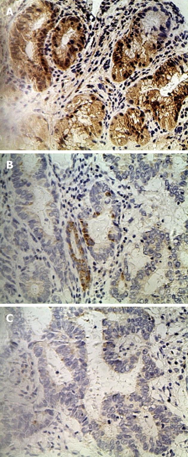
Figure 1 Immunohistochemical staining of gastric lesions using riboflavin transporter 2 gene-specific monoclonal antibodies.
A: Expression of riboflavin transporter 2 (RFT2) in normal gastric epithelium with strong staining; B: Moderate expression of RFT2 protein in gastric cancer (GC) tissue; C: Weak expression of RFT2 protein in GC tissue (original magnification, × 400).
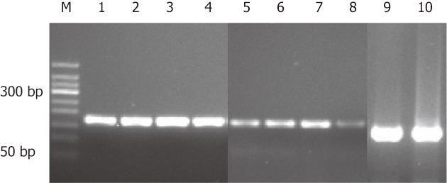
Figure 2 mRNA expression of riboflavin transporter 2 in gastric cancer tissue and control tissue.
Panels 1 to 4 and 9 are from normal gastric epithelium. Panels 5 to 8 and 10 are from gastric cancer tissues. Panels 1 to 8 show expression of riboflavin transporter 2 mRNA. Panels 9 and 10 show expression of β-actin.
Figure 3 Chromatographic profiles of a low tumor stage patient and a high tumor stage patient.
-
Citation: Eli M, Li DS, Zhang WW, Kong B, Du CS, Wumar M, Mamtimin B, Sheyhidin I, Hasim A. Decreased blood riboflavin levels are correlated with defective expression of
RFT2 gene in gastric cancer. World J Gastroenterol 2012; 18(24): 3112-3118 - URL: https://www.wjgnet.com/1007-9327/full/v18/i24/3112.htm
- DOI: https://dx.doi.org/10.3748/wjg.v18.i24.3112


