Copyright
©2012 Baishideng Publishing Group Co.
World J Gastroenterol. Jun 21, 2012; 18(23): 3008-3014
Published online Jun 21, 2012. doi: 10.3748/wjg.v18.i23.3008
Published online Jun 21, 2012. doi: 10.3748/wjg.v18.i23.3008
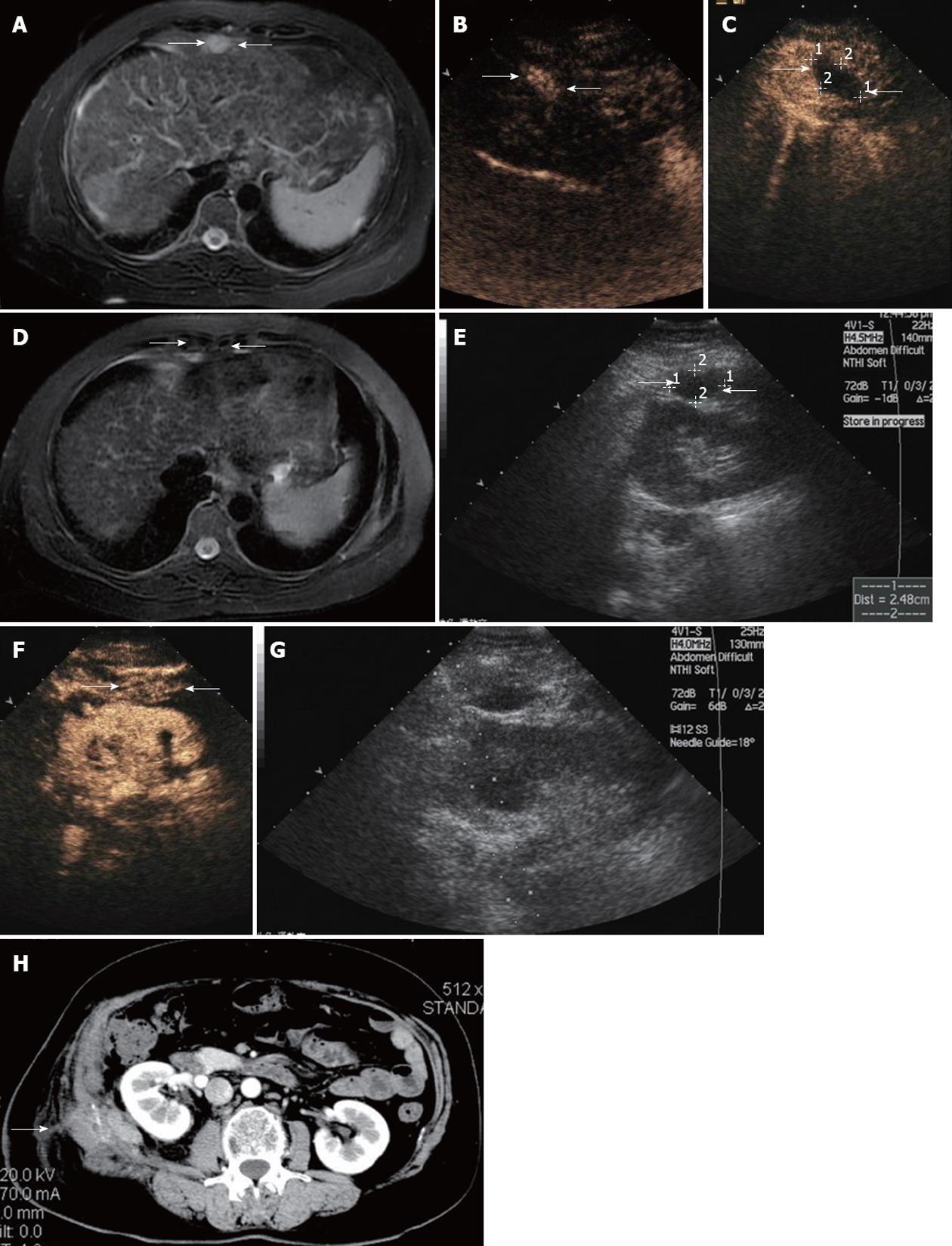
Figure 1 Ultrasound findings in a 56-year-old woman with abdominal wall tumor metastasized from liver and adrenal gland cancer.
A: Contrast-enhanced magnetic resonance imaging (MRI) scan shows a lesion with hyperenhancement in T2-weighted images in the abdominal wall (arrow); B: Arterial phase in contrast-enhanced ultrasound (CEUS) shows hyperenhancement within the lesions (arrow); C: CEUS scan obtained 3 d after microwave (MW) ablation shows a hypoechoic area with no enhancement, suggestive of complete necrosis (arrow); D: Contrast-enhanced MRI scan shows a lesion with hypoenhancement in T2-weighted phase MR image obtained 1 mo after MW ablation revealing complete ablation. Contrast-enhanced MRI scan shows a lesion with hypoenhancement in T2-weighted images in the abdominal wall (arrow); E: Sonogram obtained before MW ablation shows hypoechoic nodule of 2.48 cm in maximum diameter in the abdominal wall (arrow); F: Arterial phase in CEUS shows hyperenhancement within the lesions (arrow); G: Sonogram obtained during MW ablation shows one antenna being inserted into the nodule; H: Abdominal wall edema occurred at the right lumbar in the arterial phase of contrast-enhanced CT (arrow).
- Citation: Qi C, Yu XL, Liang P, Cheng ZG, Liu FY, Han ZY, Yu J. Ultrasound-guided microwave ablation for abdominal wall metastatic tumors: A preliminary study. World J Gastroenterol 2012; 18(23): 3008-3014
- URL: https://www.wjgnet.com/1007-9327/full/v18/i23/3008.htm
- DOI: https://dx.doi.org/10.3748/wjg.v18.i23.3008


