Copyright
©2012 Baishideng Publishing Group Co.
World J Gastroenterol. May 28, 2012; 18(20): 2582-2585
Published online May 28, 2012. doi: 10.3748/wjg.v18.i20.2582
Published online May 28, 2012. doi: 10.3748/wjg.v18.i20.2582
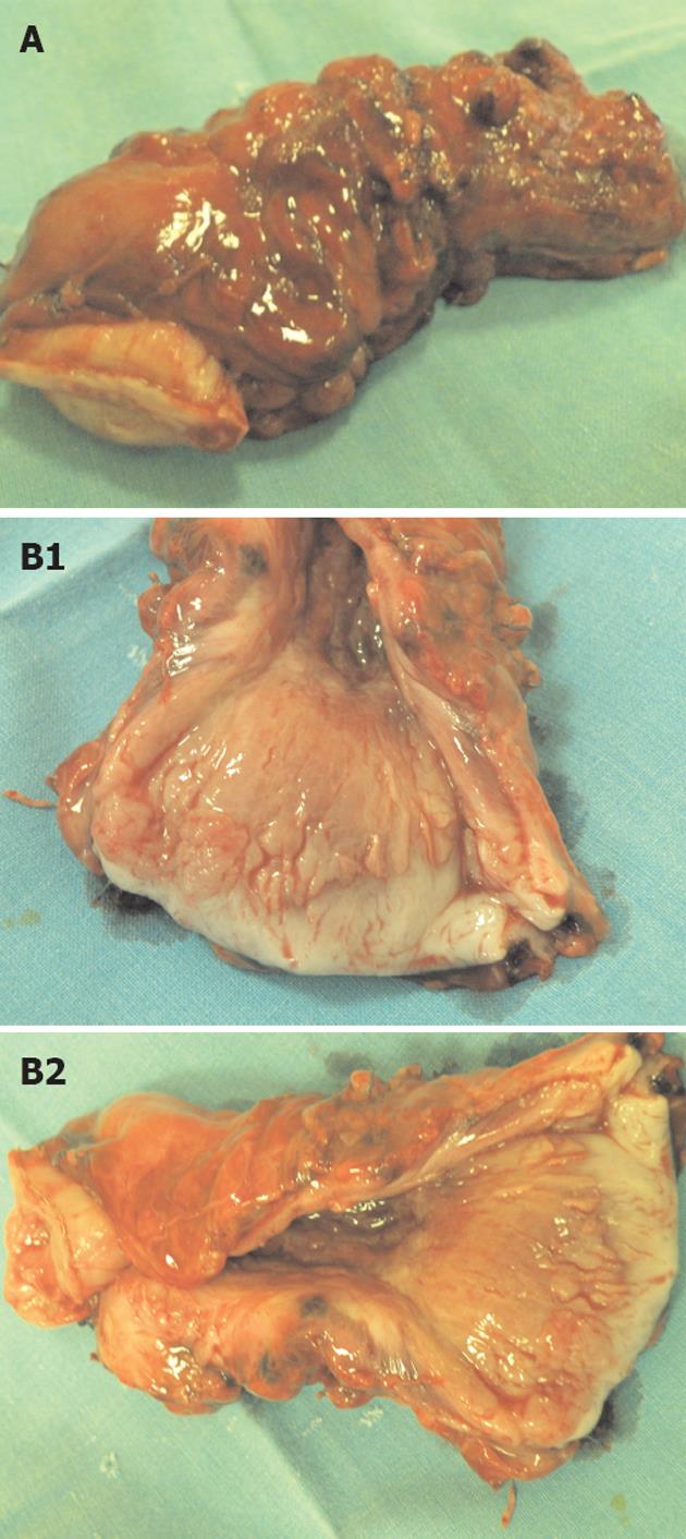
Figure 1 Pathological changes in atrophic gastritis after treatment.
A: Tubular resected stomach as a whole, 9 cm long; B1 and B2: Cross-section of stomach with its 2 cm thick wall and its atrophic mucose membrane.
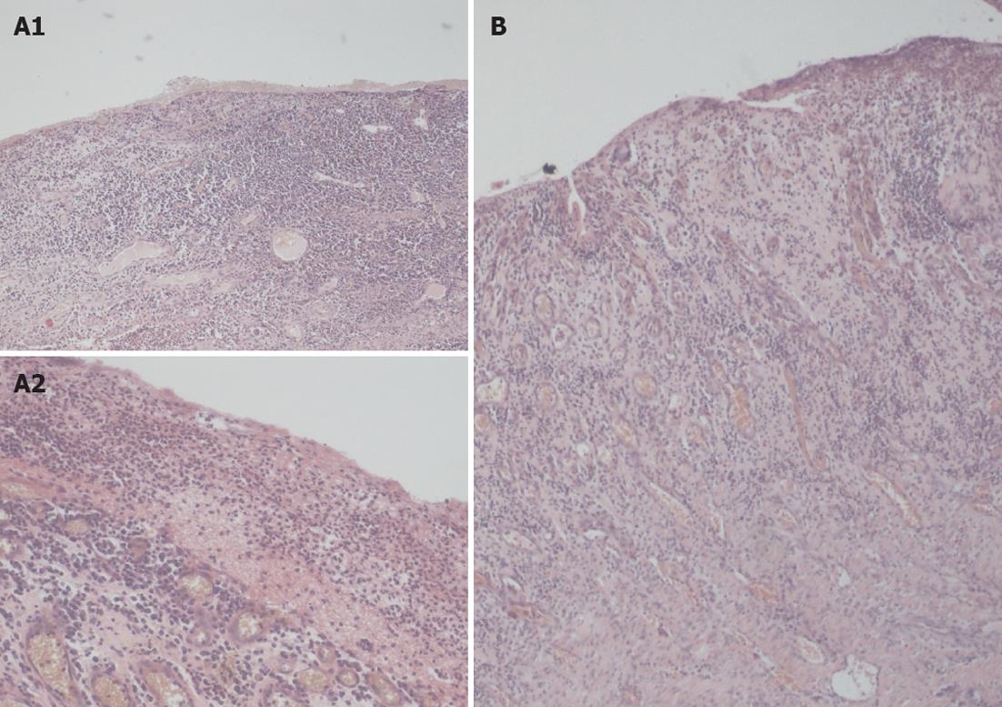
Figure 2 Representative photomicrographs of hematoxylin and eosin stained sections from the stomach’s necrosis of submucous membrane.
A: The inflammatory infiltration of the submucosal membrane of stomach (A1 magnitude 40× and A2 magnitude 100×); B: The ulceration-granulation in the stomach’s submucosal membrane, magnitude 40×.
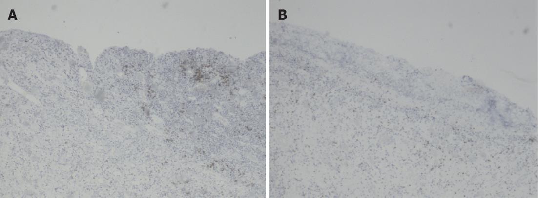
Figure 3 Representative photomicrographs sections from the stomach (magnitude 40×).
A: The large, transmural inflammatory cell infiltration of submucosal membrane CD3 (+); B: Mixed inflammatory cell infiltrations CD3 (+).
- Citation: Snarska J, Jacyna K, Janiszewski J, Shafie D, Iwanowicz K, Żurada A. Exceptionally rare cause of a total stomach resection. World J Gastroenterol 2012; 18(20): 2582-2585
- URL: https://www.wjgnet.com/1007-9327/full/v18/i20/2582.htm
- DOI: https://dx.doi.org/10.3748/wjg.v18.i20.2582


