Copyright
©2012 Baishideng Publishing Group Co.
World J Gastroenterol. May 28, 2012; 18(20): 2452-2461
Published online May 28, 2012. doi: 10.3748/wjg.v18.i20.2452
Published online May 28, 2012. doi: 10.3748/wjg.v18.i20.2452
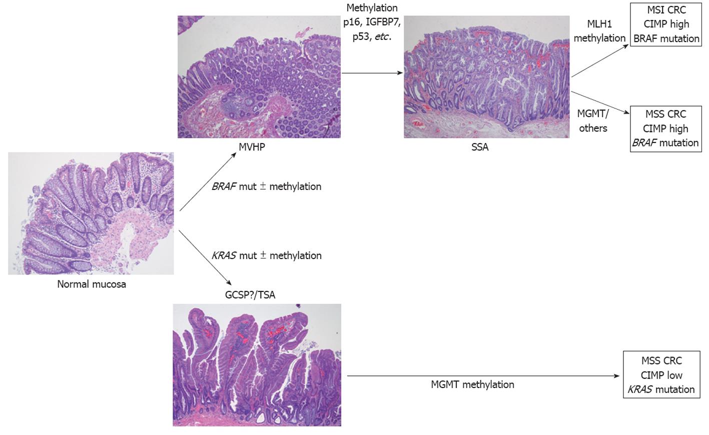
Figure 1 Model of serrated pathway of colorectal carcinogenesis.
MVHP: Microvesicular hyperplastic polyp; SSA: Sessile serrated adenoma; MGMT: Methylguanine methyltransferase; MSI: Microsatellite instability; MSS: Microsatellite stable; CRC: Colorectal cancer; CIMP: CpG island methylator phenotype; GCSP: Goblet cell serrated polyp; TSA: Traditional sessile adenomas.
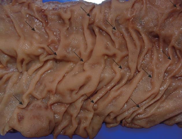
Figure 2 Segment of colectomy in a case of serrated polyposis.
Polyps are frequently small (arrows) and flat, making their endoscopic detection difficult.
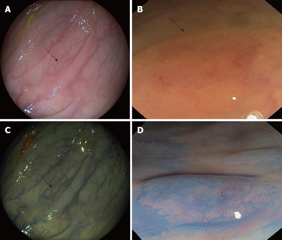
Figure 3 Endoscopic appearance of serrated polyps.
A and B: Sessile serrated adenoma (SSA) (arrows) as flat polyp on conventional optical colonoscopy; C: Narrow-band imaging appearance of polyp (arrow) seen in panel A; D: Chromoendoscopy image of SSA revealing Kudo II pattern (Images courtesy of Dr. Adolfo Parra, Hospital Central de Asturias, Oviedo, Spain).
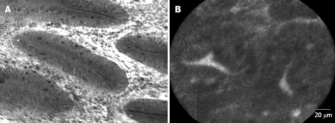
Figure 4 Confocal endomicroscopy of (A) tubular adenoma, low-grade dysplasia and (B) serrated polyp.
Both types of polyps show different shape as well as differences in the cellular structures (Images courtesy of Dr. Maria Pellisé, Hospital Clinic, Barcelona, Spain).
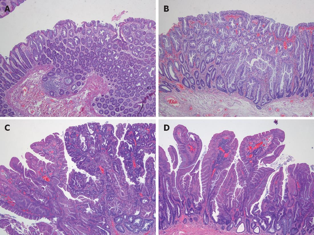
Figure 5 Pathological types of serrated polyps.
A: Microvesicular serrated polyp; B: Sessile serrated adenomas; C: Traditional serrated adenoma; D: Mixed polyp.
- Citation: Guarinos C, Sánchez-Fortún C, Rodríguez-Soler M, Alenda C, Payá A, Jover R. Serrated polyposis syndrome: Molecular, pathological and clinical aspects. World J Gastroenterol 2012; 18(20): 2452-2461
- URL: https://www.wjgnet.com/1007-9327/full/v18/i20/2452.htm
- DOI: https://dx.doi.org/10.3748/wjg.v18.i20.2452


