Copyright
©2012 Baishideng Publishing Group Co.
World J Gastroenterol. May 21, 2012; 18(19): 2438-2440
Published online May 21, 2012. doi: 10.3748/wjg.v18.i19.2438
Published online May 21, 2012. doi: 10.3748/wjg.v18.i19.2438
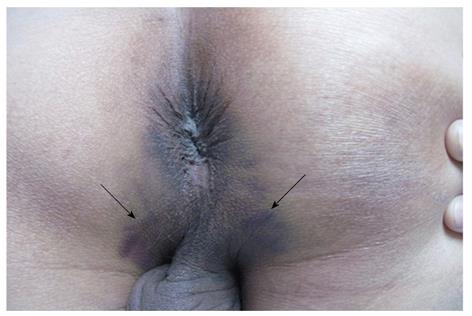
Figure 1 Pre-operative photograph of the perineum (knee-chest position) revealing widespread perianal ecchymosis (marked with black arrows).
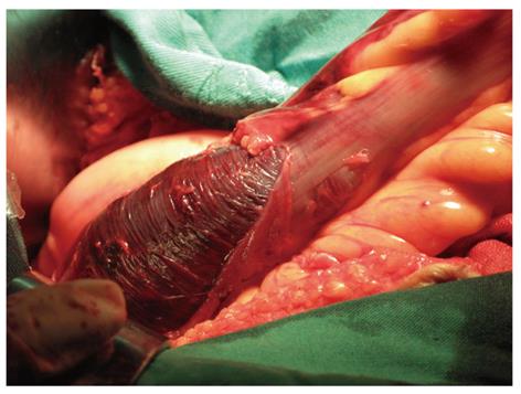
Figure 2 Intra-operative photograph of a large intramural hematoma arising beneath the rectosigmoid junction.
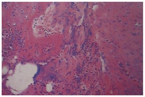
Figure 3 Extensive oozing of blood in the muscular layer of the resected rectal and fibrinoid necrosis within the vascular wall (hematoxylin and eosion stain, × 20).
- Citation: Li ZL, Wang ZJ, Han JG. Spontaneous perforation of an intramural rectal hematoma: Report of a case. World J Gastroenterol 2012; 18(19): 2438-2440
- URL: https://www.wjgnet.com/1007-9327/full/v18/i19/2438.htm
- DOI: https://dx.doi.org/10.3748/wjg.v18.i19.2438


