Copyright
©2012 Baishideng Publishing Group Co.
World J Gastroenterol. May 21, 2012; 18(19): 2320-2333
Published online May 21, 2012. doi: 10.3748/wjg.v18.i19.2320
Published online May 21, 2012. doi: 10.3748/wjg.v18.i19.2320
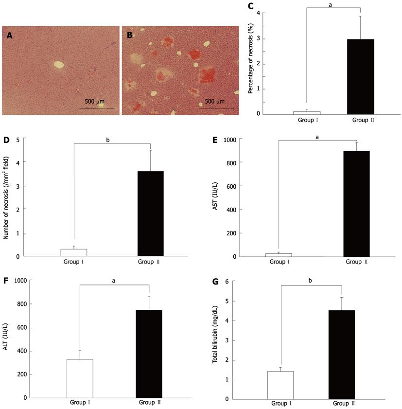
Figure 1 Histological and hematological differences between groups I and II at 6 h after 80%-partial hepatectomy.
A: Histological findings in group I mice; B: Representative images of remnant liver histology demonstrating significant multiple foci of hemorrhage and necrosis in group II mice; C: Quantitation of the size (area percentage) of hemorrhage and necrosis: Significantly larger areas and more hemorrhagic and necrotic foci were observed in group II mice than in group I mice (aP < 0.05); D: Quantitation of the number of foci of hemorrhage and necrosis per mm2: Significantly larger areas and more hemorrhagic and necrotic foci were observed in group II mice than in group I mice (bP < 0.01); E: Aspartate aminotransferase (AST) levels were significantly higher in group II mice than those in group I mice (aP < 0.05); F: Alanine aminotransaminase (ALT) levels were significantly higher in group II mice than those in group I mice (aP < 0.05); G: Total bilirubin levels were significantly higher in group II mice than those in group I mice (bP < 0.01).
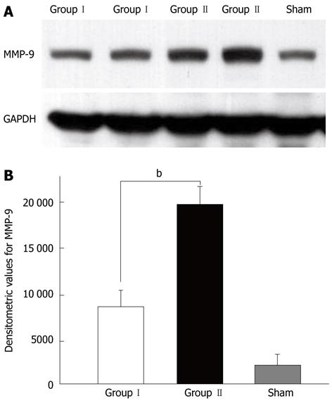
Figure 2 Western blotting analysis for matrix metalloproteinase-9 in remnant livers with (group II) and without (group I) foci of hemorrhage and necrosis after 80%-partial hepatectomy.
Representative immunoblot (A) and histogram (B) show enhanced expression of matrix metalloproteinase (MMP)-9 protein in group II mice compared with that in group I mice and sham controls (bP < 0.01).
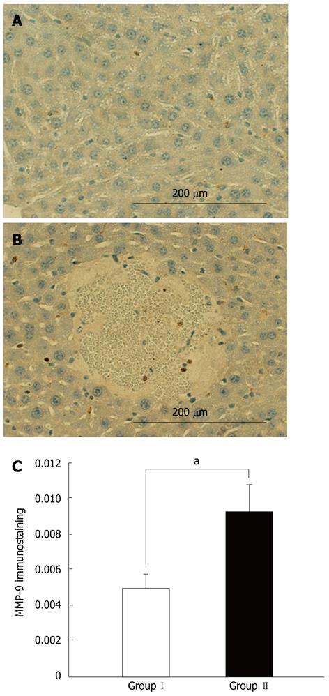
Figure 3 Immunohistochemical staining for matrix metalloproteinase-9 in the remnant liver.
A: Representative images of matrix metalloproteinase (MMP)-9 staining in a non-necrotic area in group II; B: Representative images of MMP-9 staining in a necrotic area in group II; C: Histogram of MMP-9 expression as described in the material and methods section: Enhanced MMP-9 expression was observed in areas close to the focus of necrosis (aP < 0.05).
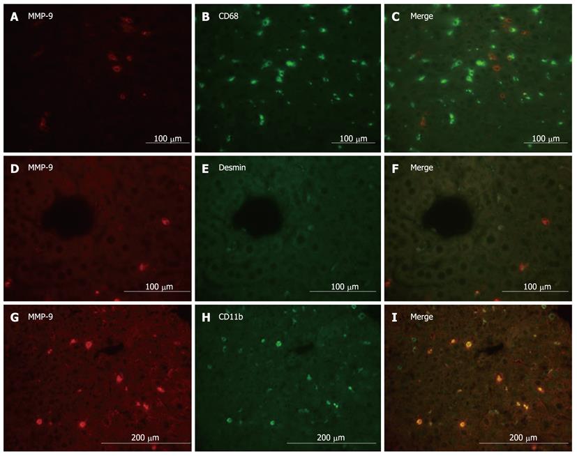
Figure 4 Co-localization analysis by dual immunofluorescence in the remnant liver after 80%-partial hepatectomy.
Co-localization analysis by dual immunofluorescence in the remnant liver for (A) matrix metalloproteinase (MMP)-9 labeled in red (Alexa Fluor 568), (B) CD68 labeled in green (Alexa Fluor 488), and (C) both MMP-9 and CD68; for (D) MMP-9 labeled in red (Alexa Fluor 568), (E) desmin labeled in green (Alexa Fluor 488), and (F) both MMP-9 and desmin; and for (G) MMP-9 labeled in red (Alexa Fluor 568), (H) CD11b labeled in green (Alexa Fluor 488), and (I) both MMP-9 and CD11b. These co-localization results demonstrate that MMP-9 protein expression is mainly localized in CD11b-positive cells, and to a lesser extent in desmin-positive cells.
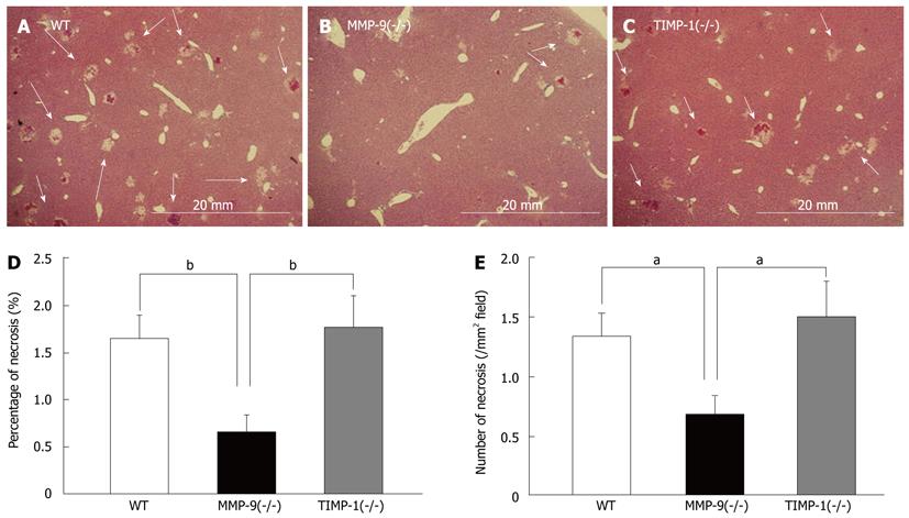
Figure 5 Histological analysis in each strain.
Histological analysis between wild-type (WT, n = 15) (A), matrix metalloproteinase (MMP)-9(-/-) (n = 15) (B), and tissue inhibitors of metalloproteinases (TIMP)-1 (-/-) mice (n = 14) (C) at 6 h after 80%-partial hepatectomy (PH). Representative images of remnant liver histology at 6 h after 80%-PH in WT, MMP-9(-/-), and TIMP-1(-/-) mice. Foci of hemorrhage and necrosis are denoted by white arrows. The percentage of the necrotic area (D) and the number of necrotic foci per mm2 (E) in remnant livers of study mice are shown. Significantly smaller and fewer necrotic foci in the remnant liver were observed in MMP-9(-/-) mice compared with WT and TIMP-1(-/-) mice (aP < 0.05, bP < 0.01).
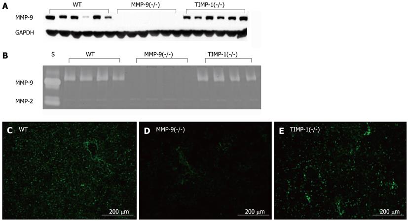
Figure 6 Expression and activity of matrix metalloproteinase-9.
Expression and activity of matrix metalloproteinase (MMP)-9 were determined using (A) western blotting analysis for MMP-9 with glyceraldehyde-3-phosphate dehydrogenase (GAPDH) as a control, (B) polyacrylamide gel electrophoresis gelatin zymography with human MMP-9 and -2 standards (S), and in situ fluorescence gelatin zymography in the remnant livers of wild-type (WT), MMP-9(-/-), and tissue inhibitors of metalloproteinases (TIMP)-1(-/-) mice at 6 h after 80%-partial hepatectomy. We observed MMP-9 protein expression and activity in WT mice (C), but they were absent in MMP-9(-/-) mice (D). Enhancement of MMP-9 protein expression and activity in TIMP-1(-/-) mice was observed (E).
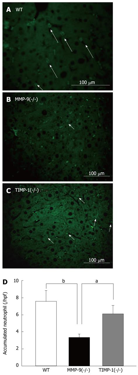
Figure 7 Immunohistochemical analysis of accumulated neutrophils with myeloperoxidase staining in remnant livers of wild-type, matrix metalloproteinase-9(-/-), and tissue inhibitors of metalloproteinases-1(-/-) mice at 6 h after 80%-partial hepatectomy.
Representative images of myeloperoxidase staining, labeled in green (Alexa Fluor 488), of cytoplasmic azurophilic granules in neutrophils (arrows) in wild-type (WT) mice in remnant liver sections in (A) WT, (B) matrix metalloproteinase (MMP)-9(-/-), and (C) tissue inhibitors of metalloproteinases (TIMP)-1(-/-) mice. A histogram shows the number of accumulated neutrophils in the remnant liver (D). Significantly fewer neutrophils were observed in the remnant liver in MMP-9(-/-) mice than in WT and TIMP-1(-/-) mice (aP < 0.05, bP < 0.01).
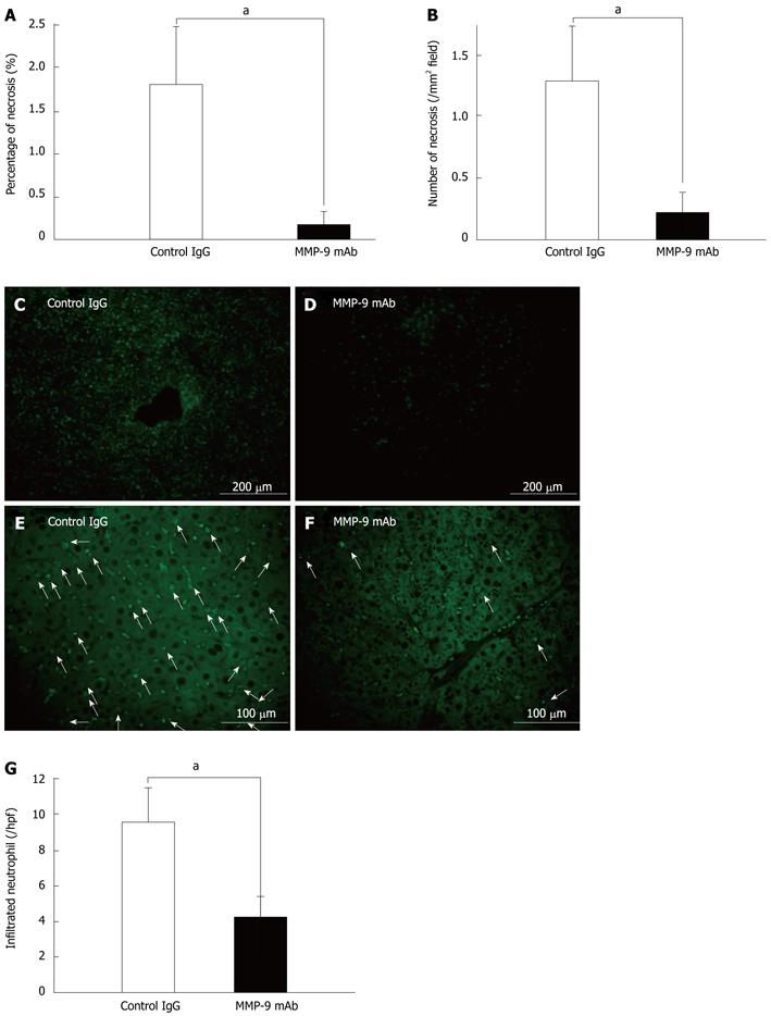
Figure 8 Effect of matrix metalloproteinase-9 monoclonal antibody on 80%-partial hepatectomy.
A, B: Treatment with matrix metalloproteinase-9 monoclonal antibody (MMP-9 mAb) in mice with 80%-partial hepatectomy (PH) resulted in a significant reduction in the size (percent area) and number of hemorrhagic and necrotic foci 6 h after 80%-PH compared with treatment using control IgG (aP < 0.05); C, D: Suppression of MMP-9 activity as determined by in situ fluorescence gelatin zymography compared with control IgG-treated mice; E, F: There was significantly less myeloperoxidase staining (arrows) in remnant liver sections in mice treated with MMP-9 mAb compared with that in control IgG-treated mice; G: A histogram shows the number of accumulated neutrophils in the remnant liver 6 h after 80%-PH. Significantly fewer neutrophils were observed in MMP-9 mAb-treated mice than in control IgG-treated animals (aP < 0.05).
- Citation: Ohashi N, Hori T, Chen F, Jermanus S, Eckman CB, Nakao A, Uemoto S, Nguyen JH. Matrix metalloproteinase-9 contributes to parenchymal hemorrhage and necrosis in the remnant liver after extended hepatectomy in mice. World J Gastroenterol 2012; 18(19): 2320-2333
- URL: https://www.wjgnet.com/1007-9327/full/v18/i19/2320.htm
- DOI: https://dx.doi.org/10.3748/wjg.v18.i19.2320


