Copyright
©2012 Baishideng Publishing Group Co.
World J Gastroenterol. May 14, 2012; 18(18): 2270-2279
Published online May 14, 2012. doi: 10.3748/wjg.v18.i18.2270
Published online May 14, 2012. doi: 10.3748/wjg.v18.i18.2270
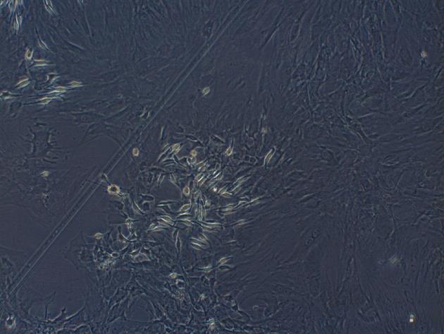
Figure 1 Third generation mesenchymal stem cells were spindle-shaped and formed spiral-like colonies (original magnification × 100).
Figure 2 The expression of mesenchymal stem cells surface markers detected by flow cytometry.
The proportion of CD29+ (A) cells was 98.6%, the proportion of CD90+ (B) cells was 99.6%, the proportion of CD45+ (C) cells was 0.89% and CD34+ (D) cells was 0.56%. B: The boundary of the cells.
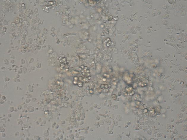
Figure 3 Separated pancreatic acinar cells (original magnification × 100).
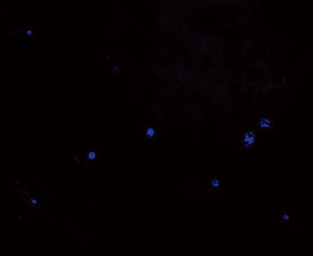
Figure 4 Transplanted mesenchymal stem cells were stained with 4,6-diamidino-2-phenylindole in advance, flash-frozen then observed under fluorescence microscope.
The blue fluorescent 4,6-diamidino-2-phenylindole-positive cells (mesenchymal stem cells) were noted in the small intestinal tissue.
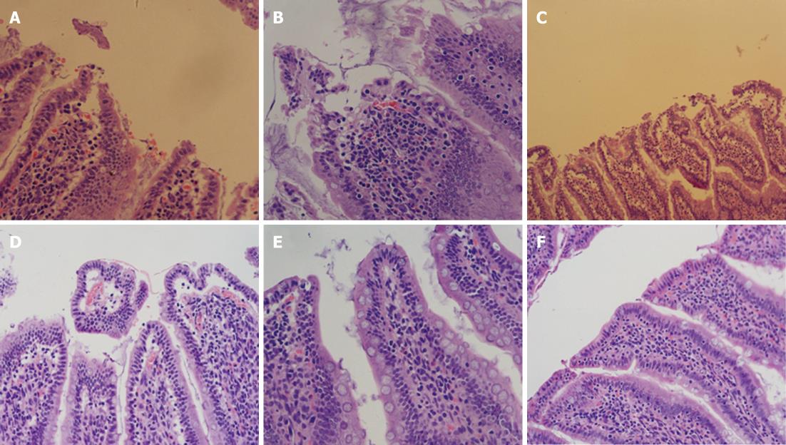
Figure 5 Description of the intestinal pathologic manifestations 6 h after mesenchymal stem cells transplantation.
A: Extensive injury of the intestinal mucosa was obvious in the severe acute pancreatitis (SAP) group; B: The dissection of the upper cortex of the intestinal mucosa was noted in the SAP + marrow mesenchymal stem cells (MSCs) group; C, D: Injury to the intestinal mucosa in the SAP (C) and SAP + MSCs groups (D) 24 h after MSCs transplantation were more severe than at 6 h; E, F: Repair of the intestinal mucosa was seen in the SAP (E) and SAP + MSCs groups (F) (HE staining, original magnification × 200).
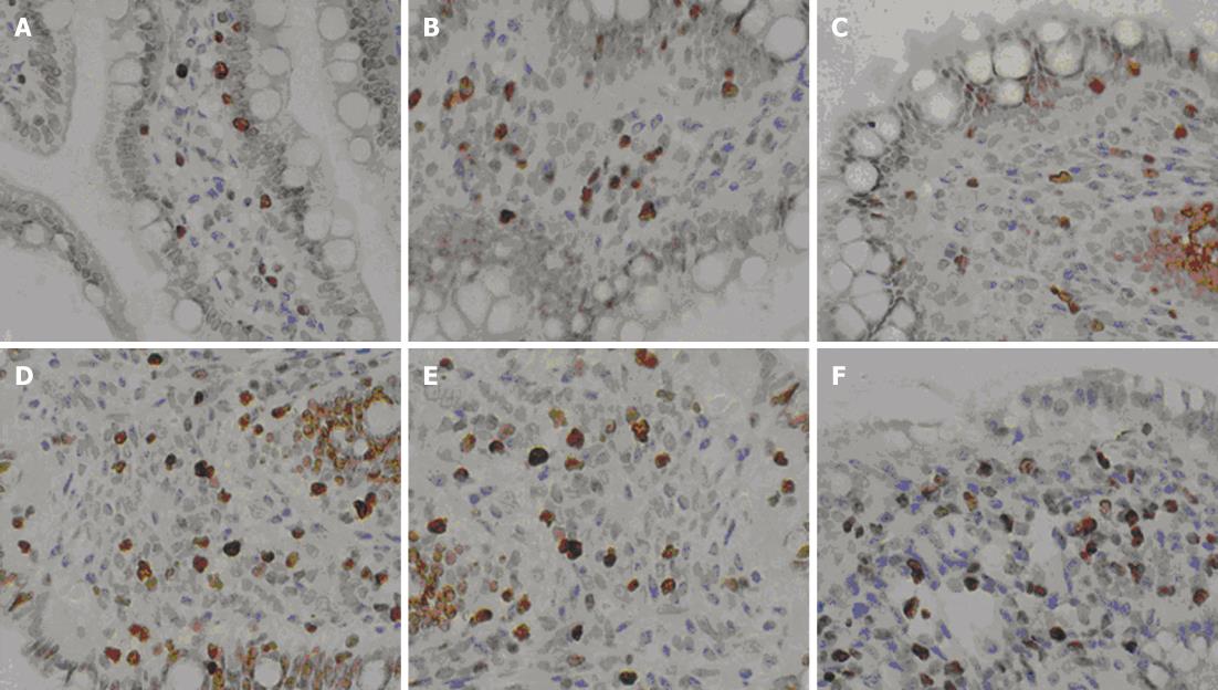
Figure 6 The immunohistochemical staining of proliferating cell nuclear antigen Ki-067 after mesenchymal stem cells transplantation at 6 h (A, B), 24 h (C, D) and 72 h (E, F).
Cell proliferation (brown cells) was obvious. The number of stained (brown) cells in the severe acute pancreatitis (SAP) + mesenchymal stem cells group (B, D, F) were significantly higher than the SAP group. Cell numbers gradually increased with time (original magnification × 400).
- Citation: Tu XH, Song JX, Xue XJ, Guo XW, Ma YX, Chen ZY, Zou ZD, Wang L. Role of bone marrow-derived mesenchymal stem cells in a rat model of severe acute pancreatitis. World J Gastroenterol 2012; 18(18): 2270-2279
- URL: https://www.wjgnet.com/1007-9327/full/v18/i18/2270.htm
- DOI: https://dx.doi.org/10.3748/wjg.v18.i18.2270


