Copyright
©2012 Baishideng Publishing Group Co.
World J Gastroenterol. May 14, 2012; 18(18): 2253-2261
Published online May 14, 2012. doi: 10.3748/wjg.v18.i18.2253
Published online May 14, 2012. doi: 10.3748/wjg.v18.i18.2253
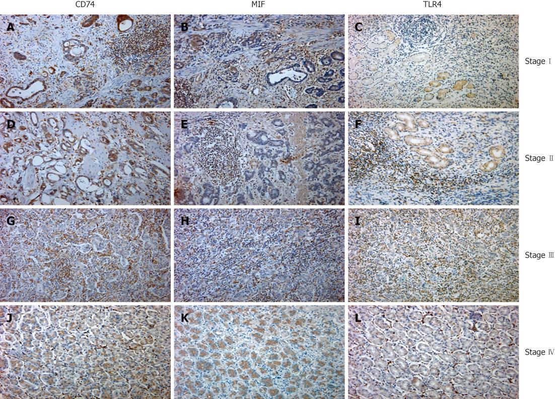
Figure 1 Representative sections show CD74, migration inhibitory factor and toll-like receptor 4 staining pattern in gastric cancer in each clinical stage.
Gastric tumor sections stained for CD74 (A, D, G and J), MIF (B, E,H and K), toll-like receptor 4 (TLR4) (C, F, I and L) in each clinical stage, and CD74, Migration inhibitory factor (MIF) and TLR4 staining for the same stage are from the same patient, demonstrating that MIF and its receptor CD74 and TLR4 are expressed in close proximity in the tumor microenvironment. Original magnifications: 200 ×.
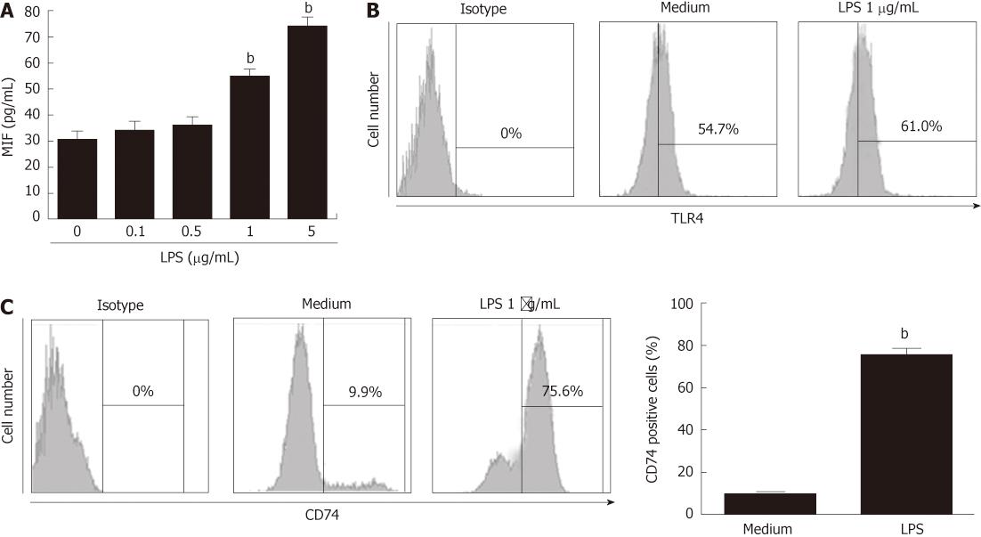
Figure 2 Lipopolysaccharide stimulation induced migration inhibitory factor and surface CD74 expression in gastric cancer cell line MKN-45.
A: MKN-45 cell line was stimulated with lipopolysaccharide (LPS) at the indicated concentration respectively for 24 h, the supernatants were collected and migration inhibitory factor (MIF) concentration was measured by enzyme-linked immunosorbent assay; B and C: MKN-45 cell line was stimulated with or without LPS (1 μg/mL) for 24 h, toll-like receptor 4 (TLR4) (B) and CD74 surface expression was detected by flow cytometry (C, left panel), the mean values of CD74-positive cells were compared between the medium and condition group (right panel). bP < 0.01 vs medium group.
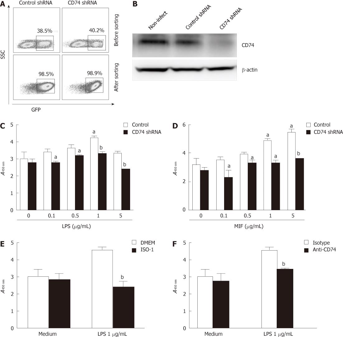
Figure 3 Lipopolysaccharide stimulation induced migration inhibitory factor and surface CD74 expression in gastric cancer cell line MKN45.
A: MKN-45 cells were transduced with control or CD74-specific shRNA, and the percentage of GFP+ cells is shown before or after flow cytometry sorting; B: CD74 expression was measured by western blotting when the MKN-45 cells were infected by control or CD74-specific shRNA; C and D: MKN-45 cells were knocked down for CD74 and stimulated with lipopolysaccharide (LPS) (C) or migration inhibitory factor (MIF) (D) for 24 h; cell proliferation was measured by CCK8; E and F: MKN-45 cells were stimulated with LPS at 1 μg/mL, and blocked with MIF antagonist ISO-1 (E) or CD74 antibody (F) for 24 h, and cell proliferation was measured by CCK8. GFP: Green fluorescent protein.aP < 0.05, bP < 0.01.
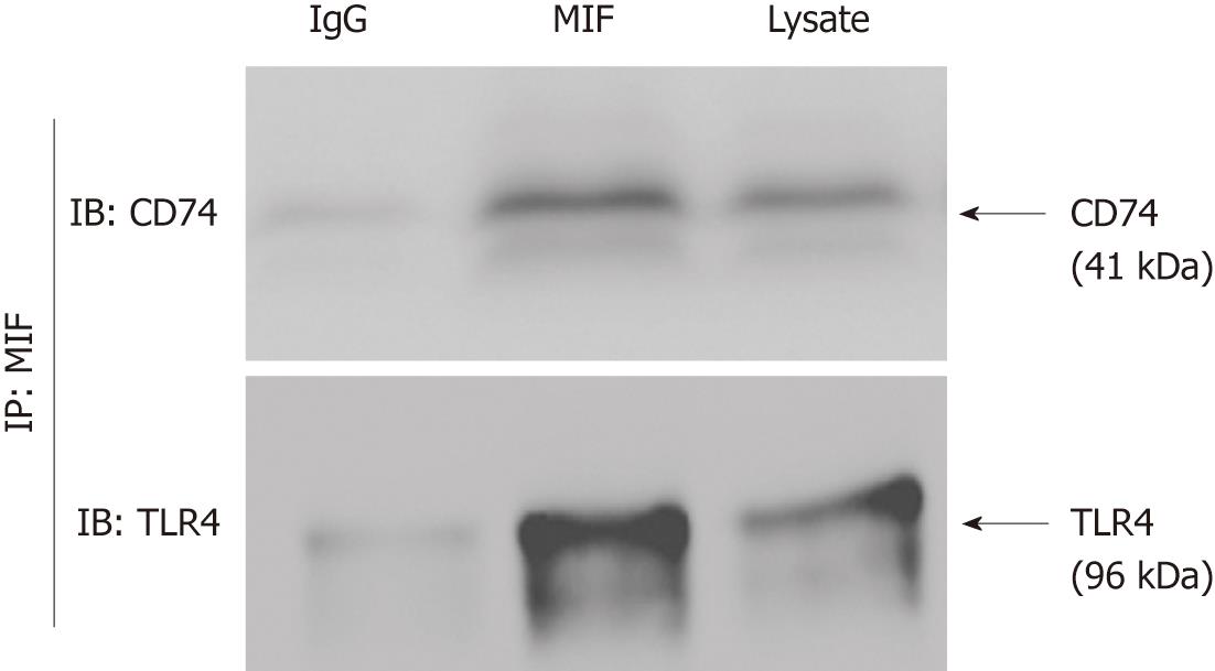
Figure 4 Migration inhibitory factor binds to CD74 and toll-like receptor 4 on gastric epithelial cells.
r-Migration inhibitory factor (MIF) was mixed with MKN-45 cell lysates and immunoprecipitated with anti-MIF with bound cell proteins. Western blotting analysis with anti-CD74 and anti-toll-like receptor 4 (TLR4). MKN-45 lysates were run as a control in the right lane, and MKN-45 cell lysates immunoprecipitated with isotype control antibody were run in the left lane.
- Citation: Zheng YX, Yang M, Rong TT, Yuan XL, Ma YH, Wang ZH, Shen LS, Cui L. CD74 and macrophage migration inhibitory factor as therapeutic targets in gastric cancer. World J Gastroenterol 2012; 18(18): 2253-2261
- URL: https://www.wjgnet.com/1007-9327/full/v18/i18/2253.htm
- DOI: https://dx.doi.org/10.3748/wjg.v18.i18.2253


